By Victoria Osinski
Lymphocytes, monocytes, and granulocytes, oh my!
There are many different immune cells (leukocytes) that are found in circulation along with other cellular components such as platelets and red blood cells. Red blood cells (RBCs) are the most abundant, followed by neutrophils and lymphocytes. The immune cells can be categorized as either mononuclear cells (monocytes and lymphocytes) or polymorphonuclear cells (granulocytes)1. A multitude of assays have been developed to isolate various cell types from the blood and they vary in complexity, cost, and cell population purity-to-yield ratios. Density gradient centrifugation is one way to isolate various peripheral blood cell types while maintaining a higher yield and lower cost. Once cells have been isolated, they can be used for a variety of applications including 1) further purification of more specific cell populations, 2) using flow cytometry to further describe and quantify cell populations, or 3) placing cells in culture to study responses to various stimulations such as pro-inflammatory cytokines .
What is density gradient centrifugation and how does it work?
Density gradient centrifugation is a method used to separate cell populations based on differential cell densities. A medium of a specified density is layered with a sample, such as whole blood, and then centrifuged. Based on the density of the medium relative to the buoyant density of the cells, certain populations will settle above, below, or in between the medium. Granulocytes (neutrophils, basophils, mast cells, and eosinophils) are the largest and densest cells, followed by monocytes, and then lymphocytes (B cells, T cells, NK cells )1,2.

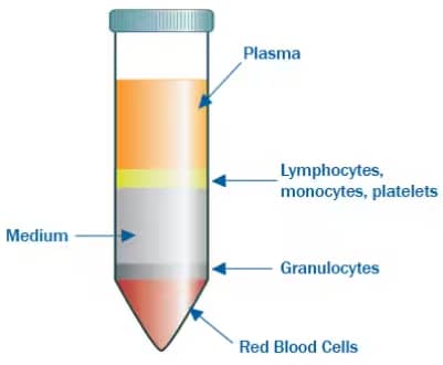
Whole blood contains various immune cells; mononuclear (monocytes and lymphocytes) and polymorphonuclear cells (granulocytes). Density gradient centrifugation allows the separation of immune cells from other whole blood components such as red blood cells, platelets and plasma.
Isolating mononuclear cells
To isolate lymphocytes, whole blood is layered upon a density gradient medium of around 1.077 g/mL. Using a medium of this density will isolate all mononuclear cells (lymphocytes and monocytes )1. Following centrifugation, the neutrophils and RBCs will be pelleted at the bottom with a layer of mononuclear cells in the density medium above. These cells can then be purified further into specific populations such as T cells or B cells.
Separating monocytes from lymphocytes
Monocytes can be further separated from lymphocytes using density gradient centrifugation because they are less dense and slightly larger. In other words, monocytes are faster to float during centrifugation in a density gradient than lymphocytes. This separation can be done with either the combination of two different density layers3 or one layer4. Following centrifugation, the monocytes should be found in a layer above the leukocytes and any other components of the whole blood that was included.
A few tips for isolating mononuclear cells:
- Avoid too much vortexing. This may activate or kill the cells.
- There may be residual RBCs following centrifugation. Use hypotonic lysis to remove these RBCs.
Isolating granulocytes
Granulocytes require a slightly different gradient protocol for isolation. Because they are generally denser cells, they require a solution with a greater density medium than that of lymphocytes and monocytes to be separated from RBCs (closer to 1.1 or 1.2 g/mL). One reliable protocol to use is a mixture of sodium metrizoate and Dextran 5005. This will produce six layers (from top to bottom): plasma, mononuclear cells, isolation media, granulocytes, more isolation media, and the red blood cell pellet. This gradient permits the separation of granulocytes and a majority of the red blood cells, and also provides the highest yield relative to other isolation methods such as microbead selection6. However, utilization of a microbead selection assay after isolating granulocytes by density gradient centrifugation may be applied to extract specific populations such as eosinophils or neutrophils. Lysis will still be required for any residual RBCs.
A few tips to reduce cell death and activation of polymorphonuclear cells during isolation:
- Don’t let the cells sit too long. These cells, in particular neutrophils, are short-lived.
- Avoid jostling the cells around too much. This means minimal vortexing (and gentle vortexing if needed) and pipetting. Tapping is a good option.
- Keep the cells at a constant temperature. Changes from cool to warm temperatures will activate the cells7,8.

Victoria (Tori) Osinski, Doctoral Candidate
University of Virginia
Victoria studies cellular mechanisms regulating vascular growth during peripheral artery disease and obesity.
-
Verhoeckx, K. et al. (2015) The impact of food bioactives on health: In vitro and Ex Vivo models. The Impact of Food Bioactives on Health: In Vitro and Ex Vivo Models.
-
Lowes, L.E. et al. (2018) Circulating Tumor Cells and Implications of the Epithelial-to-Mesenchymal Transition. Advances in Clinical Chemistry.
-
Graham, J.M. (2002) eparation of human monocytes from a leukocyte-rich plasma. TheScientificWorldJournal.
-
Graham, J.M. (2002) Separation of monocytes from whole human blood. TheScientificWorldJournal.
-
Zhou, L. et al. (2012) Impact of human granulocyte and monocyte isolation procedures on functional studies Clinical and Vaccine Immunology.
-
Forsyth, K.D. & Levinsky, R.J. (1990) Preparative procedures of cooling and re-warming increase leukocyte integrin expression and function on neotrophils. Immunological Methods.
-
Fearon, D.T. & Collins, L.A. (1983) Increased expression of C3b receptors on polymorphonuclear leukocytes induced by chemotactic factors and by purification procedures. J Immunol.