Mouse gp130 Antibody
R&D Systems, part of Bio-Techne | Catalog # MAB4681


Key Product Details
Species Reactivity
Validated:
Cited:
Applications
Validated:
Cited:
Label
Antibody Source
Product Specifications
Immunogen
Gln23-Glu617 (predicted)
Accession # Q6PDI9
Specificity
Clonality
Host
Isotype
Scientific Data Images for Mouse gp130 Antibody
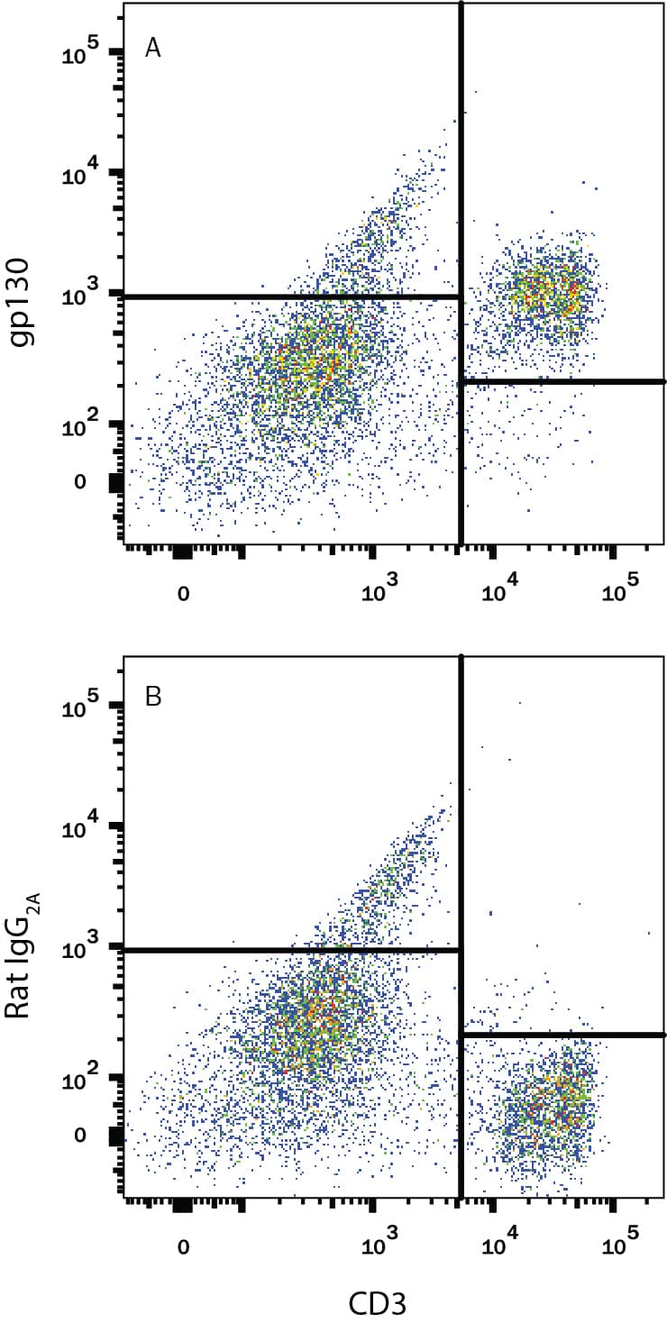
Detection of gp130 in Mouse Splenocytes by Flow Cytometry.
Mouse splenocytes were stained with Rat Anti-Mouse CD3 PE-conjugated Monoclonal Antibody (Catalog # FAB4841P) and either (A) Rat Anti-Mouse gp130 Monoclonal Antibody (Catalog # MAB4681) or (B) Rat IgG2AIsotype Control (Catalog # MAB006) followed by Allophycocyanin-conjugated Anti-Rat IgG Secondary Antibody (Catalog # F0113). View our protocol for Staining Membrane-associated Proteins.gp130 in M1 Mouse Cell Line.
gp130 was detected in immersion fixed M1 mouse myeloid leukemia cell line using Rat Anti-Mouse gp130 Monoclonal Antibody (Catalog # MAB4681) at 10 µg/mL for 3 hours at room temperature. Cells were stained using the NorthernLights™ 557-conjugated Anti-Rat IgG Secondary Antibody (red; Catalog # NL013)and counter-stained with DAPI (blue). View our protocol for Fluorescent ICC Staining of Non-adherent Cells.Detection of Mouse gp130/CD130 by Immunohistochemistry
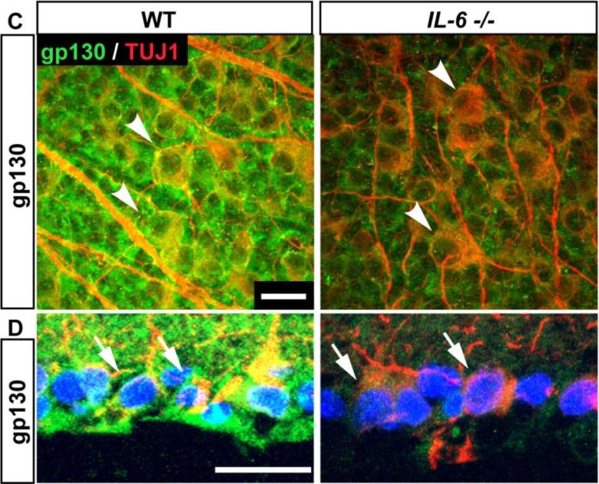
IL-6 deficiency leads to decreased gp130 protein in the retina. (A) Fold difference of Gp130 mRNA in the IL-6−/− retina normalized to WT Gp130 mRNA (dotted line) via the delta deltaCt method. (B) Densitometry analysis (right) of gp130 and beta-actin immunoblotting (left) in WT (white) and IL-6−/− (gray) retina.; student’s t-test. Error bars indicates STDEV. (C–D) Whole mount (C) and sagittal cross sections (D) of WT (left) and IL-6−/− (right) retina reveal co-localization (arrowheads; C and arrows; D) of gp130 immunolabeling (green) with beta-Tubulin+ RGCs. Error bars represent standard deviation and asterisks indicate p<0.05 (A,B). Scale bars = 20 μm (C,D). Image collected and cropped by CiteAb from the following publication (https://pubmed.ncbi.nlm.nih.gov/27747134), licensed under a CC-BY license. Not internally tested by R&D Systems.Applications for Mouse gp130 Antibody
CyTOF-ready
Flow Cytometry
Sample: Mouse splenocytes
Immunocytochemistry
Sample: Immersion fixed M1 mouse myeloid leukemia cell line
Formulation, Preparation, and Storage
Purification
Reconstitution
Formulation
Shipping
Stability & Storage
- 12 months from date of receipt, -20 to -70 °C as supplied.
- 1 month, 2 to 8 °C under sterile conditions after reconstitution.
- 6 months, -20 to -70 °C under sterile conditions after reconstitution.
Background: gp130
Gp130, the common signal transducing receptor component shared by the functional receptor complexes of the IL-6 family of cytokines, belongs to the class I cytokine receptor family. Binding of IL-6 (IL-11) to either the membrane-anchored or soluble IL-6 R (IL-11 R) initiates the association of IL-6 R (IL-11 R) with gp130 which then undergoes homo-dimerization and signal transduction. With other IL-6 family cytokines, such as LIF and OSM, signal transduction is triggered by the hetero-dimerization of gp130 and LIF R or OSM R.
Gp130 is expressed in all organs examined. Soluble gp130, which apparently arises either from proteolytic cleavage of the membrane-bound receptor or from alternative splicing, has been detected in human serum. The in vivo functions of soluble gp130 are not clearly understood. In in vitro experiments, natural or recombinant soluble gp130 has been shown to have inhibitory effects on OSM and CNTF activities.
References
- Narazaki, M. et al. (1993) Blood 82:1120.
- Taga, T. and T. Kishimoto (1997) Annu. Rev. Immunol. 15:797.
Long Name
Alternate Names
Gene Symbol
UniProt
Additional gp130 Products
Product Documents for Mouse gp130 Antibody
Product Specific Notices for Mouse gp130 Antibody
For research use only

