AKT1 [p Ser473] Antibody (17F6.B11) [Biotin]
Novus Biologicals, part of Bio-Techne | Catalog # NBP2-21678


Key Product Details
Species Reactivity
Human, Mouse, Rat, Monkey
Applications
ELISA, Flow Cytometry, Immunocytochemistry/ Immunofluorescence, Immunohistochemistry, Immunohistochemistry-Paraffin, Western Blot
Label
Biotin
Antibody Source
Monoclonal Mouse IgG1 kappa Clone # 17F6.B11
Concentration
LYOPH mg/ml
Product Specifications
Immunogen
AKT1 [p Ser473] Antibody (17F6.B11) was produced by repeated immunizations with a synthetic peptide corresponding to residues surrounding S473 of human AKT11 protein, followed by hybridoma development. (Uniprot: P31749)
Reactivity Notes
A BLAST analysis was used to suggest cross-reactivity with AKT1 pS473 from human, mouse, rat and chimpanzee sources based on 100% homology with the immunizing sequence. Cross-reactivity with AKT1 from other sources has not been determined. Cross-reactivity with AKT2 and AKT3 has not been determined.
Modification
p Ser473
Specificity
This phospho specific monoclonal antibody is specific for phosphorylated human and mouse AKT protein at S473. A BLAST analysis was used to suggest cross-reactivity with AKT pS473 from human, mouse, rat and chimpanzee sources based on 100% homology with the immunizing sequence. Cross-reactivity with AKT from other sources has not been determined. Cross-reactivity with AKT2 and AKT3 has not been determined.
Clonality
Monoclonal
Host
Mouse
Isotype
IgG1 kappa
Description
Store vial at 4C prior to restoration. For extended storage aliquot contents and freeze at -20C or below. Avoid cycles of freezing and thawing. Centrifuge product if not completely clear after standing at room temperature. This product is stable for several weeks at 4C as an undiluted liquid. Dilute only prior to immediate use.
This Mouse Monoclonal Antibody Biotin Conjugated was purified from concentrated tissue culture supernate by Protein A chromatography
This Mouse Monoclonal Antibody Biotin Conjugated was purified from concentrated tissue culture supernate by Protein A chromatography
Scientific Data Images for AKT1 [p Ser473] Antibody (17F6.B11) [Biotin]
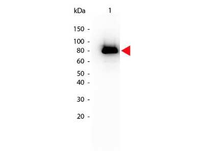
Western Blot: AKT1 [p Ser473] Antibody (17F6.B11) [Biotin] [NBP2-21678] - Western Blot of AKT1 [p Ser473] antibody (17F6.B11) [Biotin]. Lane 1: GST tagged AKT1 active recombinant protein. Lane 2: none. Load: 25 ng per lane.Primary antibody: AKT1 phospho S473 Biotin Conjugated antibody at 1:1,000 for overnight at 4C.Secondary antibody: HRP Streptavidin secondary antibody at 1:40,000 for 30 min at RT.Block for 30 min at RT.Predicted/Observed size: 79 kDa, 79 kDa for AKT1 phospho S473. Other band(s): none
Immunohistochemistry: AKT1 [p Ser473] Antibody (17F6.B11) [Biotin] [NBP2-21678] - Immunohistochemistry of AKT11 [p Ser473] antibody (17F6.B11) [Biotin]. Tissue: prostate at 40X. Fixation: FFPE buffered formalin 10% conc.Antigen retrieval: Heat, Citrate pH 6.2. Pressure Cooker, left. (pH 9 shown on right as negative control). Primary antibody: AKT1sS473 biotin 20 ug/mL for 1 h at RT.Secondary antibody: Streptavidin Conj. HRP at 10 ug/ml.Localization: nuclear and occasionally cytoplasmic.Staining: antibody as precipitated red signal with a hematoxylin purple nuclear counterstain.
Immunohistochemistry: AKT1 [p Ser473] Antibody (17F6.B11) [Biotin] [NBP2-21678] - Immunohistochemistry of AKT11 [p Ser473] antibody (17F6.B11) [Biotin]. Tissue: prostate at 40X (left) with negative control (right).Fixation: FFPE buffered formalin 10% conc.Antigen retrieval: Heat, Citrate pH 6.2. Pressure Cooker. Primary antibody: AKT1 pS473 biotin at 20 ug/mL for 1 h at RT.Secondary antibody: Streptavidin Conj. HRP at 10 ug/ml.Localization: nuclear and occasionally cytoplasmic.Staining: antibody as precipitated red signal with a hematoxylin purple nuclear counterstain.
Applications for AKT1 [p Ser473] Antibody (17F6.B11) [Biotin]
Application
Recommended Usage
ELISA
1:20000
Flow Cytometry
1:10-1:1000
Immunocytochemistry/ Immunofluorescence
1:500-1:3000
Immunohistochemistry
20 ug/mL
Immunohistochemistry-Paraffin
20 ug/mL
Western Blot
1:500-1:3000
Application Notes
This product is tested for ELISA, immunohistochemistry, immunoprecipitation and western blotting. Expect a band approximately 56 kDa in size corresponding to phosphorylated AKT protein by western blotting in the appropriate cell lysate or extract. This phospho-specific monoclonal antibody reacts with human and mouse AKT pS473 and shows minimal reactivity by ELISA against the non-phosphorylated form of the immunizing peptide. Specific conditions for reactivity should be optimized by the end user. For immunohistochemistry use formalin-fixed paraffin-embedded sections. No pre-treatment of sample is required.
Formulation, Preparation, and Storage
Purification
Protein A purified
Reconstitution
Reconstitute with 100 ul deionized water (or equivalent)
Formulation
Lyophilized from 0.02 M Potassium Phosphate, 0.15 M Sodium Chloride, pH 7.2, 10 mg/mL Bovine Serum Albumin (BSA) - Immunoglobulin and Protease free
Preservative
0.01% Sodium Azide
Concentration
LYOPH mg/ml
Shipping
The product is shipped with polar packs. Upon receipt, store it immediately at the temperature recommended below.
Stability & Storage
Store lyophilized antibody at 4C in the dark. Aliquot reconstituted liquid and store at -20C. Avoid freeze-thaw cycles.
Background: Akt1
The main function of AKT is to control inhibition of apoptosis and promote cell proliferation. Survival factors can activate AKT Ser473 and Thr308 phosphorylation sites in a transcription-independent manner, resulting in the inactivation of apoptotic signaling transduction through the tumor suppressor PTEN, an antagonist to PI3-K (5). PTEN exerts enzymatic activity as a phosphatidylinositol-3,4,5-trisphosphate (PIP3) phosphatase, opposing PI3K activity by decreasing availability of PIP3 to proliferating cells, leading to overexpression and inappropriate activation of AKT noted in many types of cancer.
AKT1 function has been linked to overall physiological growth and function (2). AKT1 has been correlated with proteus syndrome, a rare disorder characterized by overgrowth of various tissues caused by a mosaic variant in the AKT1 gene in humans.
AKT2 is strongly correlated with Type II diabetes, including phenotypes of insulin resistance, hyperglycemia and atherosclerosis (2, 6).
The function of AKT3 is specifically associated to brain development, where disruptions to AKT3 are correlated with microcephaly, hemimegalencephaly, megalencephaly and intellectual disabilities (2).
References
1. Ersahin, T., Tuncbag, N., & Cetin-Atalay, R. (2015). The PI3K/AKT/mTOR interactive pathway. Mol Biosyst, 11(7), 1946-1954. doi:10.1039/c5mb00101c
2. Cohen, M. M., Jr. (2013). The AKT genes and their roles in various disorders. Am J Med Genet A, 161a(12), 2931-2937. doi:10.1002/ajmg.a.36101
3. Georgescu, M. M. (2010). PTEN Tumor Suppressor Network in PI3K-Akt Pathway Control. Genes Cancer, 1(12), 1170-1177. doi:10.1177/1947601911407325
4. Mishra, P., Paital, B., Jena, S., Swain, S. S., Kumar, S., Yadav, M. K., . . . Samanta, L. (2019). Possible activation of NRF2 by Vitamin E/Curcumin against altered thyroid hormone induced oxidative stress via NFkB/AKT/mTOR/KEAP1 signalling in rat heart. Sci Rep, 9(1), 7408. doi:10.1038/s41598-019-43320-5
5. Wedel, S., Hudak, L., Seibel, J. M., Juengel, E., Oppermann, E., Haferkamp, A., & Blaheta, R. A. (2011). Critical analysis of simultaneous blockage of histone deacetylase and multiple receptor tyrosine kinase in the treatment of prostate cancer. Prostate, 71(7), 722-735. doi:10.1002/pros.21288
6. Rotllan, N., Chamorro-Jorganes, A., Araldi, E., Wanschel, A. C., Aryal, B., Aranda, J. F., . . . Fernandez-Hernando, C. (2015). Hematopoietic Akt2 deficiency attenuates the progression of atherosclerosis. Faseb j, 29(2), 597-610. doi:10.1096/fj.14-262097
Long Name
v-Akt Murine Thymoma Viral Oncogene Homolog 1
Alternate Names
PKB alpha, PRKBA, RAC-alpha
Gene Symbol
AKT1
UniProt
Additional Akt1 Products
Product Documents for AKT1 [p Ser473] Antibody (17F6.B11) [Biotin]
Product Specific Notices for AKT1 [p Ser473] Antibody (17F6.B11) [Biotin]
This product is for research use only and is not approved for use in humans or in clinical diagnosis. Primary Antibodies are guaranteed for 1 year from date of receipt.
Loading...
Loading...
Loading...
Loading...
Loading...
Loading...


![AKT1 [p Ser473] Antibody (17F6.B11) [Biotin] AKT1 [p Ser473] Antibody (17F6.B11) [Biotin]](https://resources.bio-techne.com/images/products/nbp2-21678_mouse-monoclonal-akt1-p-ser473-antibody-17f6-b11-biotin-235202318143164.jpg)
![AKT1 [p Ser473] Antibody (17F6.B11) [Biotin] AKT1 [p Ser473] Antibody (17F6.B11) [Biotin]](https://resources.bio-techne.com/images/products/nbp2-21678_mouse-monoclonal-akt1-p-ser473-antibody-17f6-b11-biotin-235202318143165.jpg)
![AKT1 [p Ser473] Antibody (17F6.B11) [Biotin] AKT1 [p Ser473] Antibody (17F6.B11) [Biotin]](https://resources.bio-techne.com/images/products/nbp2-21678_mouse-monoclonal-akt1-p-ser473-antibody-17f6-b11-biotin-2552023131329.jpg)