Immunohistochemistry: alpha Tubulin Antibody (DM1A) - BSA Free [NB100-690]
Immunohistochemistry: alpha Tubulin Antibody (DM1A) [NB100-690] - Analysis of formalin fixed paraffin embedded heart sections. Used at a dilution of 1:500.
Western Blot: alpha Tubulin Antibody (DM1A)BSA Free [NB100-690]
Western Blot: alpha Tubulin Antibody (DM1A) [NB100-690] - Analsis of alpha tubulin in 9 cell lysates. Lane 1. HeLa; Lane 2. JURKAT; Lane 3. COS7; Lane 4. NIH-3T3; Lane 5. PC-12; Lane 6. RAT2; Lane 7. CHO; Lane 8. MDBK; Lane 9. MDCK
Flow Cytometry: alpha Tubulin Antibody (DM1A) - BSA Free [NB100-690]
Flow Cytometry: alpha Tubulin Antibody (DM1A) [NB100-690] - Intracellular flow cytometric staining of 1 x 10^6 CHO (A) and HEK-293 (B) cells using alpha Tubulin antibody (dark blue). Isotype control shown in orange. An antibody concentration of 1 ug/1x10^6 cells was used.
Immunomicroscopy: alpha Tubulin Antibody (DM1A) - BSA Free [NB100-690]
Immunomicroscopy: alpha Tubulin Antibody (DM1A) [NB100-690] - Analysis of HeLa cells, green staining is alpha tubulin whereas red is DNA stained with propidium iodide.
Western Blot: alpha Tubulin Antibody (DM1A)BSA Free [NB100-690]
alpha-Tubulin-Antibody-DM1A-Western-Blot-NB100-690-img0054.jpg
Immunocytochemistry/ Immunofluorescence: alpha Tubulin Antibody (DM1A) - BSA Free [NB100-690]
Immunocytochemistry/Immunofluorescence: alpha Tubulin Antibody (DM1A) - BSA Free [NB100-690] - Mouse MS1 cells were fixed in 4% paraformaldehyde for 10 minutes and permeabilized in 0.05% Triton X-100 in PBS for 5 minutes. The cells were incubated with alpha Tubulin Antibody [DM1A] conjugated to Alexa Fluor 647 (NB100-690AF647) at 2 ug/ml for 1 hour at room temperature. Nuclei were counterstained with DAPI (Blue). Cells were imaged using a 100X objective and digitally deconvolved.
Immunohistochemistry-Paraffin: alpha Tubulin Antibody (DM1A) - BSA Free [NB100-690]
Immunohistochemistry-Paraffin: alpha Tubulin Antibody (DM1A) [NB100-690] - IHC analysis of a formalin fixed and paraffin embedded tissue section of mouse prostate using alpha Tubulin Antibody (DM1A) at 1:200 dilution. The signal was developed using HRP labelled secondary and DAB reagent which followed counterstaining with hematoxylin. The antibody generated a specific cytoplasmic/cytoskeletal staining in the prostate epithelial cells.
Flow Cytometry: alpha Tubulin Antibody (DM1A) - BSA Free [NB100-690]
Flow Cytometry: alpha Tubulin Antibody (DM1A) [NB100-690] - An intracellular stain was performed on HeLa cells with alpha Tubulin [DM1A] Antibody NB100-690AF700 (blue) and a matched isotype control (orange). Cells were fixed with 4% PFA and then permeabilized with 0.1% saponin. Cells were incubated in an antibody dilution of 5 ug/mL for 30 minutes at room temperature. Both antibodies were conjugated to Alexa Fluor 700.
Western Blot: alpha Tubulin Antibody (DM1A)BSA Free [NB100-690]
Western Blot: alpha Tubulin Antibody (DM1A) [NB100-690] - Western blot analysis of extracts from HeLa, COS and C6 cells using alpha Tubulin antibody (NB100-690, 1:1000, Alpha tubulin molecular weight: 50 kDa)
Western Blot: alpha Tubulin Antibody (DM1A)BSA Free [NB100-690]
Western Blot: alpha Tubulin Antibody (DM1A) [NB100-690] - Analysis of alpha tubulin (molecular weight of 50 kDa) in 9 cell lysates. Lane 1. HeLa; Lane 2. JURKAT; Lane 3. COS7; Lane 4. NIH-3T3; Lane 5. PC-12; Lane 6. RAT2; Lane 7. CHO; Lane 8. MDBK; Lane 9. MDCK
Western Blot: alpha Tubulin Antibody (DM1A)BSA Free [NB100-690]
Western Blot: alpha Tubulin Antibody (DM1A) [NB100-690] - Analysis of HeLa and COS-7 lysates. Alpha tubulin molecular weight: 50 kDa.
Immunocytochemistry/ Immunofluorescence: alpha Tubulin Antibody (DM1A) - BSA Free [NB100-690]
Immunocytochemistry/Immunofluorescence: alpha Tubulin Antibody (DM1A) [NB100-690] - IF Confocal analysis of C6 cells using alpha Tubulin antibody (NB100-690, 1:50). An Alexa Fluor 488-conjugated Goat to mouse IgG was used as secondary antibody (green, A). Actin filaments were labeled with Alexa Fluor 568 phalloidin (red, B). DAPI was used to stain the cell nuclei (blue, C).
Immunocytochemistry/ Immunofluorescence: alpha Tubulin Antibody (DM1A) - BSA Free [NB100-690]
Immunocytochemistry/Immunofluorescence: alpha Tubulin Antibody (DM1A) [NB100-690] - HeLa cells were fixed for 10 minutes using 10% formalin and then permeabilized for 5 minutes using 1X TBS + 0.5% Triton-X100. The cells were incubated with anti-alpha Tubulin (DM1A) (NB100-690) at a 1:200 dilution overnight at 4C and detected with an anti-mouse Dylight 488 (Green) at a 1:500 dilution. Nuclei were counterstained with DAPI (Blue). Cells were imaged using a 40X objective.
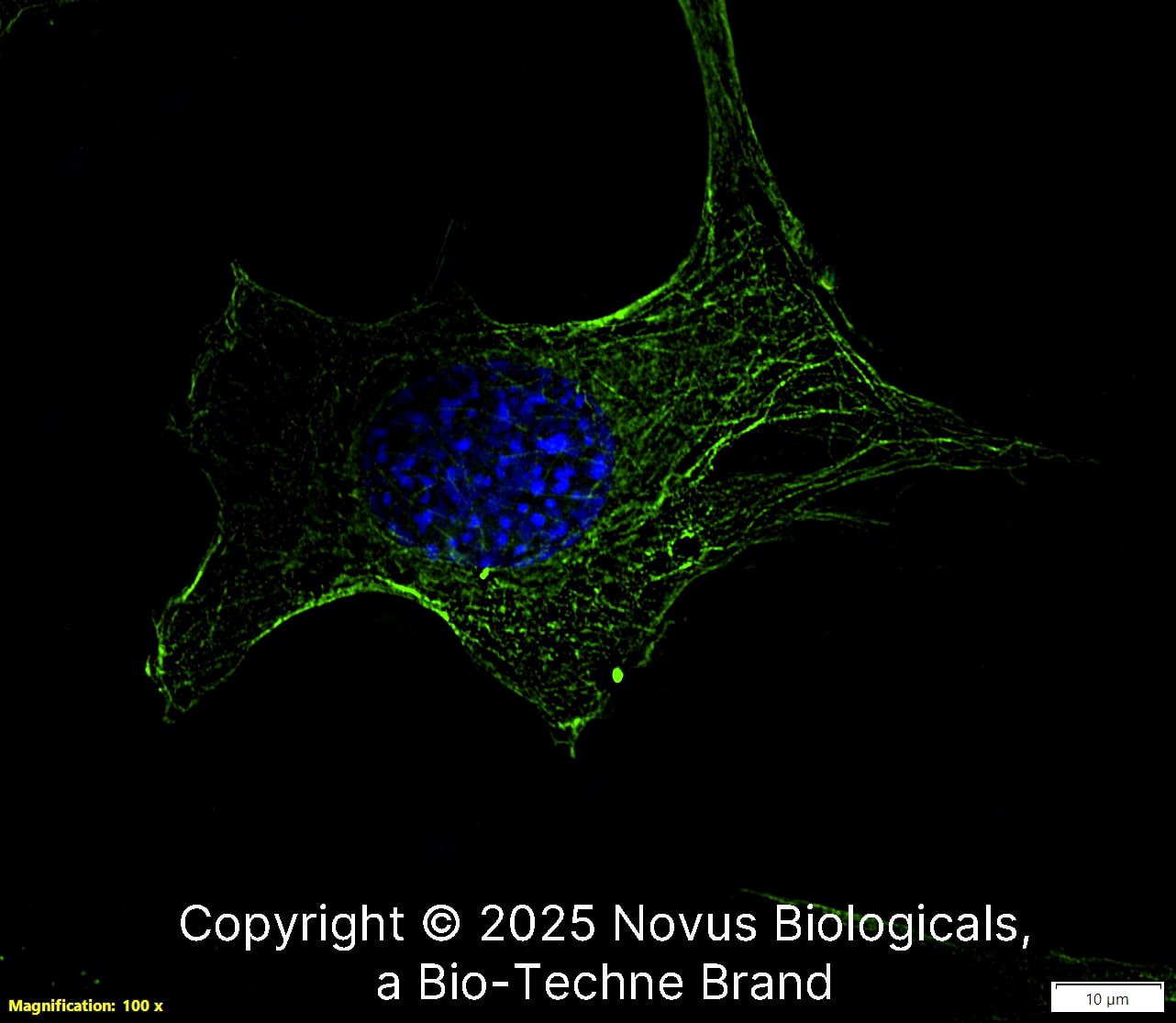
Immunocytochemistry/ Immunofluorescence: alpha Tubulin Antibody (DM1A) - BSA Free [NB100-690]
Immunocytochemistry/Immunofluorescence: alpha Tubulin Antibody (DM1A) [NB100-690] - Staining of skin fibroblasts.
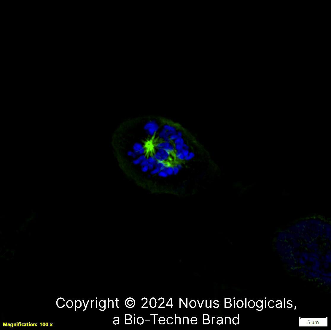
Immunocytochemistry/ Immunofluorescence: alpha Tubulin Antibody (DM1A) - BSA Free [NB100-690]
Immunocytochemistry/Immunofluorescence: alpha Tubulin Antibody (DM1A) [NB100-690] - Analysis of embryonic fibroblasts in the anaphase portion of mitosis.
Immunocytochemistry/ Immunofluorescence: alpha Tubulin Antibody (DM1A) - BSA Free [NB100-690]
Immunocytochemistry/Immunofluorescence: alpha Tubulin Antibody (DM1A) [NB100-690] - U-251 MG cells were fixed in 4% paraformaldehyde for 10 minutes and permeabilized in 0.05% Triton X-100 in PBS for 5 minutes. The cells were incubated with anti-alpha Tubulin Antibody [DM1A] conjugated to Alexa Fluor 488 (NB100-690AF488) at 5 ug/ml for 1 hour at room temperature. Nuclei were counterstained with DAPI (Blue). Cells were imaged using a 100X objective and digitally deconvolved.
Immunocytochemistry/ Immunofluorescence: alpha Tubulin Antibody (DM1A) - BSA Free [NB100-690]
Immunocytochemistry/Immunofluorescence: alpha Tubulin Antibody (DM1A) [NB100-690] - A431 cells were fixed in 4% paraformaldehyde for 10 minutes and permeabilized in 0.05% Triton X-100 in PBS for 5 minutes. The cells were incubated with anti-alpha Tubulin Antibody [DM1A] conjugated to Alexa Fluor 488 (NB100-690AF488) at 5 ug/ml for 1 hour at room temperature. Nuclei were counterstained with DAPI (Blue). Cells were imaged using a 100X objective and digitally deconvolved.
Immunocytochemistry/ Immunofluorescence: alpha Tubulin Antibody (DM1A) - BSA Free [NB100-690]
Immunocytochemistry/Immunofluorescence: alpha Tubulin Antibody (DM1A) [NB100-690] - NIH3T3 cells were fixed in 4% paraformaldehyde for 10 minutes and permeabilized in 0.05% Triton X-100 in PBS for 5 minutes. The cells were incubated with anti-alpha Tubulin Antibody [DM1A] conjugated to Alexa Fluor 488 (NB100-690AF488) at 5 ug/ml for 1 hour at room temperature. Nuclei were counterstained with DAPI (Blue). Cells were imaged using a 100X objective and digitally deconvolved.
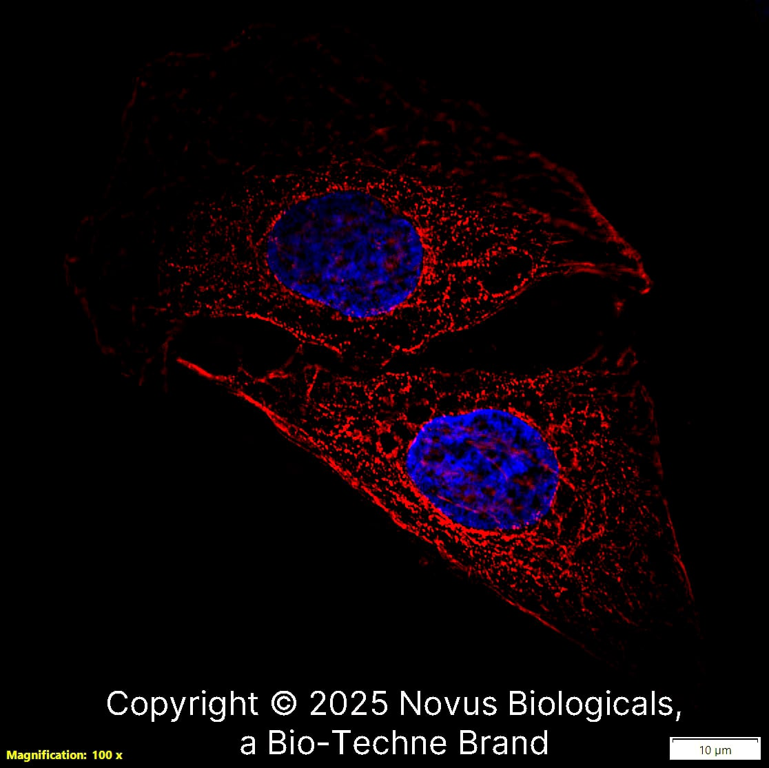
Immunocytochemistry/ Immunofluorescence: alpha Tubulin Antibody (DM1A) - BSA Free [NB100-690]
Immunocytochemistry/Immunofluorescence: alpha Tubulin Antibody (DM1A) [NB100-690] - HeLa cells were fixed in 4% paraformaldehyde for 10 minutes and permeabilized in 0.05% Triton X-100 in PBS for 5 minutes. The cells were incubated with alpha Tubulin Antibody [DM1A] conjugated to Janelia Fluor 549 (NB100-690JF549) at 5 ug/ml for 1 hour at room temperature. Nuclei were counterstained with DAPI (Blue). Cells were imaged using a 100X objective and digitally deconvolved.
Immunohistochemistry: alpha Tubulin Antibody (DM1A) - BSA Free [NB100-690]
Immunohistochemistry: alpha Tubulin Antibody (DM1A) [NB100-690] - Analysis of paraffin embedded colon sections.
Immunohistochemistry: alpha Tubulin Antibody (DM1A) - BSA Free [NB100-690]
Immunohistochemistry: alpha Tubulin Antibody (DM1A) [NB100-690] - Analysis of small intestine tissue fixed with formalin and paraffin embedded showing cytoplasmic and cytoskeletal staining of glandular cells.
Immunohistochemistry-Paraffin: alpha Tubulin Antibody (DM1A) - BSA Free [NB100-690]
Immunohistochemistry-Paraffin: alpha Tubulin Antibody (DM1A) [NB100-690] - IHC analysis of a formalin fixed paraffin embedded tissue section of mouse skeletal muscle using alpha Tubulin Antibody (DM1A) at 1:100 dilution with HRP-DAB detection and hematoxylin counterstaining. The antibody generated a strong cytoplasmic signal in the muscle cells with cytoplasmic-nuclear signal in the endothelial cells.
Immunohistochemistry-Paraffin: alpha Tubulin Antibody (DM1A) - BSA Free [NB100-690]
Immunohistochemistry-Paraffin: alpha Tubulin Antibody (DM1A) [NB100-690] - IHC analysis of a formalin fixed paraffin embedded tissue section of mouse lung using alpha Tubulin Antibody (DM1A) at 1:100 dilution with HRP-DAB detection and hematoxylin counterstaining. The antibody generated chunks of cytoplasmic signal in the alveolar and bronchiolar epithelial cells.
Immunohistochemistry-Paraffin: alpha Tubulin Antibody (DM1A) - BSA Free [NB100-690]
Immunohistochemistry-Paraffin: alpha Tubulin Antibody (DM1A) [NB100-690] - IHC analysis of a formalin fixed paraffin embedded tissue section of mouse heart using alpha Tubulin Antibody (DM1A) at 1:100 dilution with HRP-DAB detection and hematoxylin counterstaining. The antibody generated a strong and specific cytoplasmic signal in the muscle cells.
Flow Cytometry: alpha Tubulin Antibody (DM1A) - BSA Free [NB100-690]
Flow Cytometry: alpha Tubulin Antibody (DM1A) [NB100-690] - Analysis of PE conjugate of NB100-690. An intracellular stain was performed on RAW 246.7 cells with Alpha Tubulin antibody (DM1A) NB100-690PE (blue) and a matched isotype control NBP2-27287PE (orange). Cells were fixed with 4% PFA and then permeablized wi
Flow Cytometry: alpha Tubulin Antibody (DM1A) - BSA Free [NB100-690]
Flow Cytometry: alpha Tubulin Antibody (DM1A) [NB100-690] - Analysis of PE conjugate of NB100-690. An intracellular stain was performed on SH-SY5Y cells with Alpha Tubulin antibody (DM1A) NB100-690PE (blue) and a matched isotype control NBP2-27287PE (orange). Cells were fixed with 4% PFA and then permeablized with
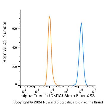
Flow (Intracellular): alpha Tubulin Antibody (DM1A) - BSA Free [NB100-690]
Flow (Intracellular): alpha Tubulin Antibody (DM1A) [NB100-690] - An intracellular stain was performed on HeLa cells with alpha Tubulin Antibody (DM1A) NB100-690AF488 (blue) and a matched isotype control (orange). Cells were fixed with 4% PFA and then permeablized with 0.1% saponin. Cells were incubated in an antibody dilution of 5 ug/mL for 30 minutes at room temperature. Both antibodies were conjugated to Alexa Fluor 488. Image from the Alexa Fluor 488 version of this antibody.
Flow Cytometry: alpha Tubulin Antibody (DM1A) - BSA Free [NB100-690]
Flow Cytometry: alpha Tubulin Antibody (DM1A) [NB100-690] - An intracellular stain was performed on HeLa cells with alpha Tubulin (DM1A) Antibody NB100-690G (blue) and a matched isotype control (orange). Cells were fixed with 4% PFA and then permeabilized with 0.1% saponin. Cells were incubated in an antibody dilution of 5 ug/mL for 30 minutes at room temperature. Both antibodies were conjugated to DyLight 488.
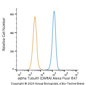
Flow Cytometry: alpha Tubulin Antibody (DM1A) - BSA Free [NB100-690]
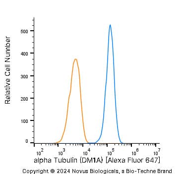
Flow Cytometry: alpha Tubulin Antibody (DM1A) [NB100-690] - An intracellular stain was performed on HeLa cells with alpha Tubulin [DM1A] Antibody NB100-690AF647 (blue) and a matched isotype control (orange). Cells were fixed with 4% PFA and then permeabilized with 0.1% saponin. Cells were incubated in an antibody dilution of 2.5 ug/mL for 30 minutes at room temperature. Both antibodies were conjugated to Alexa Fluor 647.
Flow Cytometry: alpha Tubulin Antibody (DM1A) - BSA Free [NB100-690]
Flow Cytometry: alpha Tubulin Antibody (DM1A) [NB100-690] - An intracellular stain was performed on HeLa cells with alpha Tubulin (DM1A) Antibody NB100-690JF646 (blue) and a matched isotype control (orange). Cells were fixed with 4% PFA and then permeabilized with 0.1% saponin. Cells were incubated in an antibody dilution of 2.5 ug/mL for 30 minutes at room temperature. Both antibodies were conjugated to Janelia Fluor 646.
Immunomicroscopy: alpha Tubulin Antibody (DM1A) - BSA Free [NB100-690]
Immunomicroscopy: alpha Tubulin Antibody (DM1A) [NB100-690] - Staining of the marine parasite Cryptocaryon irritans mouth. Large bundles of microtubules form a cytophyrigeal basket.
Western Blot: alpha Tubulin Antibody (DM1A) - BSA Free [NB100-690] -
GS treatment increases markers of beiging in 3T3-L1 adipocytes. GS treatment upregulates markers of beiging, including UCP1 (A), TBX1 (B), and beta-3AR (C) proteins. Data presented as mean ± SEM from n = 4 replicates per group. * p < 0.05, *** p < 0.001 vs. control. Abbreviations: isoproterenol (ISO), uncoupling protein 1 (UCP1), glyceraldehyde 3-phosphate dehydrogenase (GAPDH), T-box protein 1 (TBX1), beta-3 adrenergic receptor ( beta-3AR).
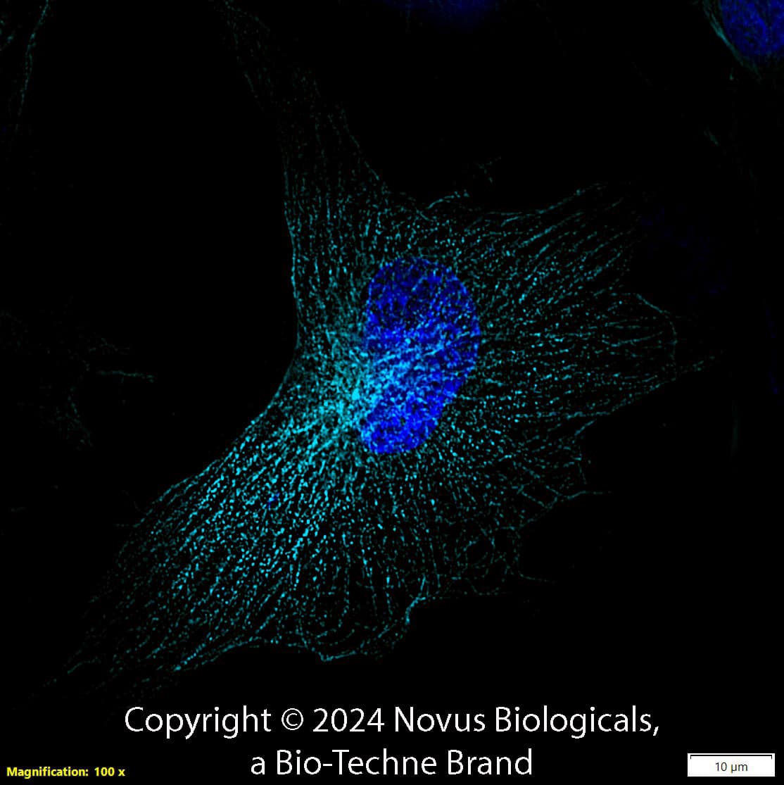
Alpha Tubulin (DM1A) in U-251 MG Human Cell Line -
Alpha Tubulin (DM1A) was detected in immersion fixed U-251 MG human glioblastoma cell line using Mouse anti-alpha Tubulin (DM1A) Protein-G purified Monoclonal Antibody conjugated to Alexa Fluor® 647 (Catalog # NB100-690AF647) (light blue) at 2 µg/mL overnight at 4C. Cells were counterstained with DAPI (dark blue). Cells were imaged using 100X objective and digitally deconvolved.
Immunohistochemistry-Paraffin: Mouse Monoclonal alpha Tubulin Antibody (DM1A) [IMG-80196] [NB100-690]
Immunohistochemistry-Paraffin: Mouse Monoclonal alpha Tubulin Antibody (DM1A) [IMG-80196] [NB100-690] - Immunofluorescence staining of human tonsil FFPE tissue in a dilution of 1:50 (Catalog #
NB100-690AF488) in 3% BSA with overnight incubation at 4°C. Heat mediated antigen retrieval at pH 9. Image from a verified customer review.
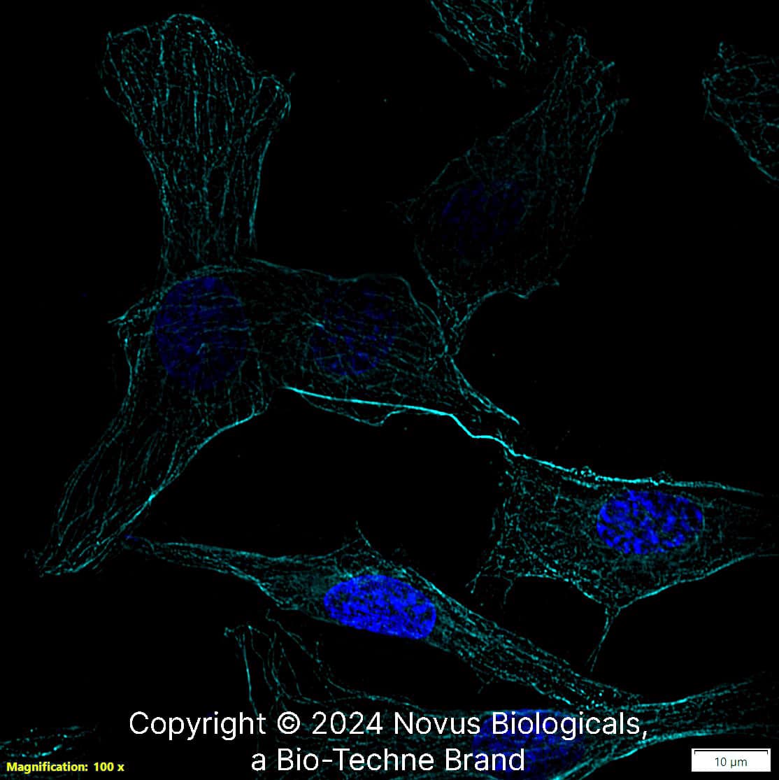
Alpha Tubulin (DM1A) in U-251 MG Human Cell Line -
Alpha Tubulin (DM1A) was detected in immersion fixed U-251 MG human glioblastoma cell line using Mouse anti-alpha Tubulin (DM1A) Protein-G purified Monoclonal Antibody conjugated to Alexa Fluor® 647 (Catalog # NB100-690AF647) (light blue) at 2 µg/mL overnight at 4C. Cells were counterstained with DAPI (dark blue). Cells were imaged using 100X objective and digitally deconvolved.
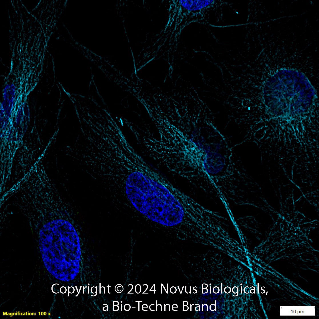
alpha Tubulin (DM1A) in FR Rat Cell Line.
alpha Tubulin (DM1A) was detected in immersion FR rat skin fibroblast cell line using Mouse anti-alpha Tubulin (DM1A) Protein G Purified Monoclonal Antibody conjugated to Alexa Fluor® 647 (Catalog # NB100-690AF647) (light blue) at 2 µg/mL overnight at 4C. Cells were counterstained with DAPI (dark blue). Cells were imaged using a 100X objective and digitally deconvolved.
Detection of alpha Tubulin (DM1A) in U-251 MG Human Cell Line by Flow Cytometry.
U-251 MG human glioblastoma cell line was stained with Mouse anti-alpha Tubulin (DM1A) Protein-G purified Monoclonal Antibody conjugated to Alexa Fluor® 647 (Catalog # NB100-690AF647, blue histogram) or matched control antibody (orange histogram).
Western Blot: alpha Tubulin Antibody (DM1A) - BSA Free [NB100-690] -
Western Blot: alpha Tubulin Antibody (DM1A) - BSA Free [NB100-690] - MiR-375-3p negatively regulates Derlin-1 & blocks EMT in BFTC909 cells. (A) Western blot revealed the restoration of Derlin-1, MMP-2, Snail, & ZEB1 after co-transfection of miR-375-3p mimics & CMV-Derlin-1 compared with cells transfected with miR-375-3p alone in BFTC909 cells with alpha-tubulin as a reference (B) Quantification of the protein levels of Derlin-1, occludin, MMP-2, Snail, & ZEB1 from (A) (N = 3). (C) miR-375-3p suppressed BFTC909 cell migration ability but restored by Derlin-1 overexpression (N = 3). (D) miR-375-3p repressed invasion of BFTC909 cells but restored by Derlin-1 overexpression (N = 3). Data were represented as mean ± SD; * p < 0.05, ** p < 0.01. Image collected & cropped by CiteAb from the following publication (https://pubmed.ncbi.nlm.nih.gov/35205628), licensed under a CC-BY license. Not internally tested by Novus Biologicals.
Western Blot: alpha Tubulin Antibody (DM1A) - BSA Free [NB100-690] -
Western Blot: alpha Tubulin Antibody (DM1A) - BSA Free [NB100-690] - Nifedipine stimulated tremendous production of reactive oxygen species (ROS), & KIM-1 in 24 & 48 h. (a) Nifedipine 30 µM-treated group had induced a higher ROS (3.3-fold vs. control, p < 0.01) compared to H2O2 500 μM. (2.7-fold vs. control, p < 0.01). (b,c) Nifedipine 7.5, 15, & 30 μM-treated groups for 24 h (tubulin as internal control) had upregulated KIM-1 in dose dependent fashion (101%, 102%, p < 0.05, & 122%, p < 0.01 respectively) & reduced to 86%, 91%, & 80% in 48 h (actin as internal control), respectively. p-values ≤ 0.05 (marked as *) were considered statistically significant. In addition, p-values ≤ 0.01 are marked as **. Image collected & cropped by CiteAb from the following publication (https://pubmed.ncbi.nlm.nih.gov/30934807), licensed under a CC-BY license. Not internally tested by Novus Biologicals.
Western Blot: alpha Tubulin Antibody (DM1A) - BSA Free [NB100-690] -
Western Blot: alpha Tubulin Antibody (DM1A) - BSA Free [NB100-690] - Pre-treatment with 0.5 mM sodium arsenite (SA) enhances permissivity in a cell-type-specific manner across reovirus strains. (A) CV-1, HeLa, L929, or HPDE cells were left untreated (no SA) or were treated with 0.5 mM SA for 30 min prior to infection (Pre-SA). Following this, cells were infected with T3D such that ~20% to 50% of cells were infected & at 18 h p.i. cells were fixed & immunostained for μNS & DAPI to visualize viral factories (VFs). The percent of cells containing VFs was quantified ((# of cells containing VFs/total # of cells) × 100) from three independent experiments. The expression level of μNS (B) & μ1 (C) was determined in CV-1, L929, or HeLa cells either left untreated (no SA) or treated with 0.5 mM SA for 30 min (Pre-SA) before infection with T3D at MOI = 1. At 18 h p.i., cells were harvested & the expression level of the indicated proteins was determined by immunoblot. M = mock. Densitometry analysis of the band intensity for μNS & μ1 was adjusted to the matched alpha-tubulin loading control for two independent experiments. Columns represent mean ± SEM. (D) CV-1; (E) L929; or (F) HeLa cells were left untreated (no SA) or were treated with 0.5 mM SA prior to infection (Pre-SA). Cells were then infected with the reovirus strains, T3D, T1L, or T3A, as described in (A). At 18 h p.i., cells were fixed & immunostained for μNS & DAPI to detect VFs. The percent of cells containing VFs was quantified ((# of cells containing VFs/total # of cells) × 100) from at least two independent experiments. * p < 0.05; ** p < 0.01; two-tailed unpaired t test. The error bars indicate S.D. Image collected & cropped by CiteAb from the following publication (https://pubmed.ncbi.nlm.nih.gov/31216693), licensed under a CC-BY license. Not internally tested by Novus Biologicals.
Western Blot: alpha Tubulin Antibody (DM1A) - BSA Free [NB100-690] -
Western Blot: alpha Tubulin Antibody (DM1A) - BSA Free [NB100-690] - Simvastatin increases cytotoxicity in lung cancer cells. (A) Relative survival (%) in lung cancer cells treated with simvastatin for 48 h using 3-[4,5-dimethylthiazol-2-yl]-2,5 diphenyl tetrazolium bromide (MTT) assays is shown. (B) The half-maximal inhibitory concentration (IC50) of simvastatin is summarized, & western blots of p53 in low-invasive CL1-0 & high-invasive Bm7 cells are shown with elongation factor 1 alpha (EF1 alpha) used as a loading control. (C) Z score of statins as well as simvastatin in lung cancer cell lines from NCI-DTP database, z score > 0 for sensitive & <0 resistant. (D) Apoptotic H1299 (null p53), A549 (wild type p53), Bm7-shGFP (mutant p53), & Bm7-shTP53 (knock-down p53) cells treated with simvastatin were detected using flow cytometry, *P < 0.05 & **P < 0.01. (E) Apoptotic HCC827-shGFP (mutant p53) & HCC827-shTP53 (knock-down p53) cells treated with simvastatin & cisplatin were detected using flow cytometry, *P < 0.05 & **P < 0.01. (F) Western blots of indicated proteins involved in apoptosis & autophagy in both Bm7 & HCC827 cells with control (shGFP) & p53 knockdown (shTP53) treated with simvastatin is shown. MDM2, murine double minute 2; AKT, serine–threonine kinase; PARP, poly (ADP-ribose) polymerase; mTOR, mammalian target of rapamycin; WT, wild type. Full-length blots/gels are presented in Supplementary Fig. 1. Image collected & cropped by CiteAb from the following publication (https://pubmed.ncbi.nlm.nih.gov/31892709), licensed under a CC-BY license. Not internally tested by Novus Biologicals.
Western Blot: alpha Tubulin Antibody (DM1A) - BSA Free [NB100-690] -
Western Blot: alpha Tubulin Antibody (DM1A) - BSA Free [NB100-690] - Pre-treatment with 0.5 mM sodium arsenite (SA) enhances permissivity in a cell-type-specific manner across reovirus strains. (A) CV-1, HeLa, L929, or HPDE cells were left untreated (no SA) or were treated with 0.5 mM SA for 30 min prior to infection (Pre-SA). Following this, cells were infected with T3D such that ~20% to 50% of cells were infected & at 18 h p.i. cells were fixed & immunostained for μNS & DAPI to visualize viral factories (VFs). The percent of cells containing VFs was quantified ((# of cells containing VFs/total # of cells) × 100) from three independent experiments. The expression level of μNS (B) & μ1 (C) was determined in CV-1, L929, or HeLa cells either left untreated (no SA) or treated with 0.5 mM SA for 30 min (Pre-SA) before infection with T3D at MOI = 1. At 18 h p.i., cells were harvested & the expression level of the indicated proteins was determined by immunoblot. M = mock. Densitometry analysis of the band intensity for μNS & μ1 was adjusted to the matched alpha-tubulin loading control for two independent experiments. Columns represent mean ± SEM. (D) CV-1; (E) L929; or (F) HeLa cells were left untreated (no SA) or were treated with 0.5 mM SA prior to infection (Pre-SA). Cells were then infected with the reovirus strains, T3D, T1L, or T3A, as described in (A). At 18 h p.i., cells were fixed & immunostained for μNS & DAPI to detect VFs. The percent of cells containing VFs was quantified ((# of cells containing VFs/total # of cells) × 100) from at least two independent experiments. * p < 0.05; ** p < 0.01; two-tailed unpaired t test. The error bars indicate S.D. Image collected & cropped by CiteAb from the following publication (https://pubmed.ncbi.nlm.nih.gov/31216693), licensed under a CC-BY license. Not internally tested by Novus Biologicals.
Alpha Tubulin (DM1A) in U-2 OS Human Cell Line.
Alpha Tubulin (DM1A) was detected in immersion fixed U-2 OS human osteosarcoma cell line using Mouse anti-Alpha Tubulin (DM1A) Protein G Purified Monoclonal Antibody conjugated to Alexa Fluor ® 488 (Catalog # NB100-690AF488) (green) at 2 µg/mL overnight at 4C. Cells were counterstained with DAPI (dark blue). Cells were imaged using a 100X objective and digitally deconvolved.
Detection of alpha Tubulin (DM1A) in U-2 OS Human Cell Line by Flow Cytometry.
An intracellular stain was performed on U-2 OS human osteosarcoma cell line with Mouse anti-alpha Tubulin (DM1A) Protein-G purified Monoclonal Antibody conjugated to Alexa Fluor ® 488 (Catalog # NB100-690AF488, blue histogram) or matched control antibody (orange histogram) at 5 µg/mL for 30 minutes at RT.
Detection of alpha Tubulin (DM1A) in A431 Human Cell Line by Flow Cytometry.
An intracellular stain was performed on A431 human skin carcinoma cell line using Mouse anti-alpha Tubulin (DM1A) Protein-G purified Monoclonal Antibody conjugated to Alexa Fluor ® 647 (Catalog # NB100-690AF647, blue histogram) or matched control antibody (orange histogram) at 2.5 µg/mL for 30 minutes at RT.
Alpha Tubulin (DM1A) in NIH-3T3 Mouse Cell Line.
Alpha Tubulin (DM1A) was detected in immersion fixed NIH-3T3 Mouse fibroblast cell line using Mouse anti-Alpha Tubulin (DM1A) Protein G Purified Monoclonal Antibody conjugated to Alexa Fluor® 488 (Catalog # NB100-690AF488) (green) at 2 µg/mL overnight at 4C. Cells were counterstained with DAPI (dark blue). Cells were imaged using a 100X objective and digitally deconvolved.
Alpha Tubulin (DM1A) in U-2 OS Human Cell Line.
Alpha Tubulin (DM1A) was detected in immersion fixed U-2 OS human osteosarcoma cell line using Mouse anti-Alpha Tubulin (DM1A) Protein G Purified Monoclonal Antibody conjugated to Biotin (Catalog # NB100-690B) at 2 µg/mL overnight at 4C. Cells were stained using Streptavidin conjugated to DyLight 550 (red) and counterstained with DAPI (blue). Cells were imaged using a 100X objective and digitally deconvolved.

![Simple Western: alpha Tubulin Antibody (DM1A)BSA Free [NB100-690] Simple Western: alpha Tubulin Antibody (DM1A)BSA Free [NB100-690]](https://resources.bio-techne.com/images/products/alpha-Tubulin-Antibody-DM1A-Simple-Western-NB100-690-img0023.jpg)
![Immunohistochemistry: alpha Tubulin Antibody (DM1A) - BSA Free [NB100-690] Immunohistochemistry: alpha Tubulin Antibody (DM1A) - BSA Free [NB100-690]](https://resources.bio-techne.com/images/products/alpha-Tubulin-Antibody-DM1A-Immunohistochemistry-NB100-690-img0028.jpg)
![Immunohistochemistry: alpha Tubulin Antibody (DM1A) - BSA Free [NB100-690] Immunohistochemistry: alpha Tubulin Antibody (DM1A) - BSA Free [NB100-690]](https://resources.bio-techne.com/images/products/alpha-Tubulin-Antibody-DM1A-Immunohistochemistry-NB100-690-img0029.jpg)
![Immunohistochemistry: alpha Tubulin Antibody (DM1A) - BSA Free [NB100-690] Immunohistochemistry: alpha Tubulin Antibody (DM1A) - BSA Free [NB100-690]](https://resources.bio-techne.com/images/products/alpha-Tubulin-Antibody-DM1A-Immunohistochemistry-NB100-690-img0031.jpg)
![Western Blot: alpha Tubulin Antibody (DM1A)BSA Free [NB100-690] Western Blot: alpha Tubulin Antibody (DM1A)BSA Free [NB100-690]](https://resources.bio-techne.com/images/products/alpha-Tubulin-Antibody-DM1A-Western-Blot-NB100-690-img0032.jpg)
![Flow Cytometry: alpha Tubulin Antibody (DM1A) - BSA Free [NB100-690] Flow Cytometry: alpha Tubulin Antibody (DM1A) - BSA Free [NB100-690]](https://resources.bio-techne.com/images/products/alpha-Tubulin-Antibody-DM1A-Flow-Cytometry-NB100-690-img0016.jpg)
![Immunomicroscopy: alpha Tubulin Antibody (DM1A) - BSA Free [NB100-690] Immunomicroscopy: alpha Tubulin Antibody (DM1A) - BSA Free [NB100-690]](https://resources.bio-techne.com/images/products/alpha-Tubulin-Antibody-DM1A-Immunomicroscopy-NB100-690-img0037.jpg)
![Western Blot: alpha Tubulin Antibody (DM1A)BSA Free [NB100-690] Western Blot: alpha Tubulin Antibody (DM1A)BSA Free [NB100-690]](https://resources.bio-techne.com/images/products/alpha-Tubulin-Antibody-DM1A-Western-Blot-NB100-690-img0054.jpg)
![Immunocytochemistry/ Immunofluorescence: alpha Tubulin Antibody (DM1A) - BSA Free [NB100-690] Immunocytochemistry/ Immunofluorescence: alpha Tubulin Antibody (DM1A) - BSA Free [NB100-690]](https://resources.bio-techne.com/images/products/alpha-Tubulin-Antibody-DM1A-BSA-Free-Immunocytochemistry-Immunofluorescence-NB100-690-img0061.jpg)
![Immunohistochemistry-Paraffin: alpha Tubulin Antibody (DM1A) - BSA Free [NB100-690] Immunohistochemistry-Paraffin: alpha Tubulin Antibody (DM1A) - BSA Free [NB100-690]](https://resources.bio-techne.com/images/products/alpha-Tubulin-Antibody-DM1A-Immunohistochemistry-Paraffin-NB100-690-img0044.jpg)
![Flow Cytometry: alpha Tubulin Antibody (DM1A) - BSA Free [NB100-690] Flow Cytometry: alpha Tubulin Antibody (DM1A) - BSA Free [NB100-690]](https://resources.bio-techne.com/images/products/alpha-Tubulin-Antibody-DM1A-Flow-Cytometry-NB100-690-img0060.jpg)
![Western Blot: alpha Tubulin Antibody (DM1A)BSA Free [NB100-690] Western Blot: alpha Tubulin Antibody (DM1A)BSA Free [NB100-690]](https://resources.bio-techne.com/images/products/alpha-Tubulin-Antibody-DM1A-Western-Blot-NB100-690-img0019.jpg)
![Western Blot: alpha Tubulin Antibody (DM1A)BSA Free [NB100-690] Western Blot: alpha Tubulin Antibody (DM1A)BSA Free [NB100-690]](https://resources.bio-techne.com/images/products/alpha-Tubulin-Antibody-DM1A-Western-Blot-NB100-690-img0025.jpg)
![Western Blot: alpha Tubulin Antibody (DM1A)BSA Free [NB100-690] Western Blot: alpha Tubulin Antibody (DM1A)BSA Free [NB100-690]](https://resources.bio-techne.com/images/products/alpha-Tubulin-Antibody-DM1A-Western-Blot-NB100-690-img0035.jpg)
![Immunocytochemistry/ Immunofluorescence: alpha Tubulin Antibody (DM1A) - BSA Free [NB100-690] Immunocytochemistry/ Immunofluorescence: alpha Tubulin Antibody (DM1A) - BSA Free [NB100-690]](https://resources.bio-techne.com/images/products/alpha-Tubulin-Antibody-DM1A-Immunocytochemistry-Immunofluorescence-NB100-690-img0018.jpg)
![Immunocytochemistry/ Immunofluorescence: alpha Tubulin Antibody (DM1A) - BSA Free [NB100-690] Immunocytochemistry/ Immunofluorescence: alpha Tubulin Antibody (DM1A) - BSA Free [NB100-690]](https://resources.bio-techne.com/images/products/alpha-Tubulin-Antibody-DM1A-Immunocytochemistry-Immunofluorescence-NB100-690-img0042.jpg)
![Immunocytochemistry/ Immunofluorescence: alpha Tubulin Antibody (DM1A) - BSA Free [NB100-690] Immunocytochemistry/ Immunofluorescence: alpha Tubulin Antibody (DM1A) - BSA Free [NB100-690]](https://resources.bio-techne.com/images/products/alpha-Tubulin-Antibody-DM1A-Immunocytochemistry-Immunofluorescence-NB100-690-img0030.jpg)
![Immunocytochemistry/ Immunofluorescence: alpha Tubulin Antibody (DM1A) - BSA Free [NB100-690] Immunocytochemistry/ Immunofluorescence: alpha Tubulin Antibody (DM1A) - BSA Free [NB100-690]](https://resources.bio-techne.com/images/products/alpha-Tubulin-Antibody-DM1A-Immunocytochemistry-Immunofluorescence-NB100-690-img0033.jpg)
![Immunocytochemistry/ Immunofluorescence: alpha Tubulin Antibody (DM1A) - BSA Free [NB100-690] Immunocytochemistry/ Immunofluorescence: alpha Tubulin Antibody (DM1A) - BSA Free [NB100-690]](https://resources.bio-techne.com/images/products/alpha-Tubulin-Antibody-DM1A-Immunocytochemistry-Immunofluorescence-NB100-690-img0055.jpg)
![Immunocytochemistry/ Immunofluorescence: alpha Tubulin Antibody (DM1A) - BSA Free [NB100-690] Immunocytochemistry/ Immunofluorescence: alpha Tubulin Antibody (DM1A) - BSA Free [NB100-690]](https://resources.bio-techne.com/images/products/alpha-Tubulin-Antibody-DM1A-Immunocytochemistry-Immunofluorescence-NB100-690-img0056.jpg)
![Immunocytochemistry/ Immunofluorescence: alpha Tubulin Antibody (DM1A) - BSA Free [NB100-690] Immunocytochemistry/ Immunofluorescence: alpha Tubulin Antibody (DM1A) - BSA Free [NB100-690]](https://resources.bio-techne.com/images/products/alpha-Tubulin-Antibody-DM1A-Immunocytochemistry-Immunofluorescence-NB100-690-img0057.jpg)
![Immunocytochemistry/ Immunofluorescence: alpha Tubulin Antibody (DM1A) - BSA Free [NB100-690] Immunocytochemistry/ Immunofluorescence: alpha Tubulin Antibody (DM1A) - BSA Free [NB100-690]](https://resources.bio-techne.com/images/products/alpha-Tubulin-Antibody-DM1A-Immunocytochemistry-Immunofluorescence-NB100-690-img0058.jpg)
![Immunocytochemistry/ Immunofluorescence: alpha Tubulin Antibody (DM1A) - BSA Free [NB100-690] Immunocytochemistry/ Immunofluorescence: alpha Tubulin Antibody (DM1A) - BSA Free [NB100-690]](https://resources.bio-techne.com/images/products/alpha-Tubulin-Antibody-DM1A-Immunocytochemistry-Immunofluorescence-NB100-690-img0059.jpg)
![Immunohistochemistry: alpha Tubulin Antibody (DM1A) - BSA Free [NB100-690] Immunohistochemistry: alpha Tubulin Antibody (DM1A) - BSA Free [NB100-690]](https://resources.bio-techne.com/images/products/alpha-Tubulin-Antibody-DM1A-Immunohistochemistry-NB100-690-img0036.jpg)
![Immunohistochemistry: alpha Tubulin Antibody (DM1A) - BSA Free [NB100-690] Immunohistochemistry: alpha Tubulin Antibody (DM1A) - BSA Free [NB100-690]](https://resources.bio-techne.com/images/products/alpha-Tubulin-Antibody-DM1A-Immunohistochemistry-NB100-690-img0038.jpg)
![Immunohistochemistry-Paraffin: alpha Tubulin Antibody (DM1A) - BSA Free [NB100-690] Immunohistochemistry-Paraffin: alpha Tubulin Antibody (DM1A) - BSA Free [NB100-690]](https://resources.bio-techne.com/images/products/alpha-Tubulin-Antibody-DM1A-Immunohistochemistry-Paraffin-NB100-690-img0039.jpg)
![Immunohistochemistry-Paraffin: alpha Tubulin Antibody (DM1A) - BSA Free [NB100-690] Immunohistochemistry-Paraffin: alpha Tubulin Antibody (DM1A) - BSA Free [NB100-690]](https://resources.bio-techne.com/images/products/alpha-Tubulin-Antibody-DM1A-Immunohistochemistry-Paraffin-NB100-690-img0040.jpg)
![Immunohistochemistry-Paraffin: alpha Tubulin Antibody (DM1A) - BSA Free [NB100-690] Immunohistochemistry-Paraffin: alpha Tubulin Antibody (DM1A) - BSA Free [NB100-690]](https://resources.bio-techne.com/images/products/alpha-Tubulin-Antibody-DM1A-Immunohistochemistry-Paraffin-NB100-690-img0041.jpg)
![Flow Cytometry: alpha Tubulin Antibody (DM1A) - BSA Free [NB100-690] Flow Cytometry: alpha Tubulin Antibody (DM1A) - BSA Free [NB100-690]](https://resources.bio-techne.com/images/products/alpha-Tubulin-Antibody-DM1A-Flow-Cytometry-NB100-690-img0045.jpg)
![Flow Cytometry: alpha Tubulin Antibody (DM1A) - BSA Free [NB100-690] Flow Cytometry: alpha Tubulin Antibody (DM1A) - BSA Free [NB100-690]](https://resources.bio-techne.com/images/products/alpha-Tubulin-Antibody-DM1A-Flow-Cytometry-NB100-690-img0046.jpg)
![Flow (Intracellular): alpha Tubulin Antibody (DM1A) - BSA Free [NB100-690] Flow (Intracellular): alpha Tubulin Antibody (DM1A) - BSA Free [NB100-690]](https://resources.bio-techne.com/images/products/alpha-Tubulin-Antibody-DM1A-Flow-Intracellular-NB100-690-img0049.jpg)
![Flow Cytometry: alpha Tubulin Antibody (DM1A) - BSA Free [NB100-690] Flow Cytometry: alpha Tubulin Antibody (DM1A) - BSA Free [NB100-690]](https://resources.bio-techne.com/images/products/alpha-Tubulin-Antibody-DM1A-Flow-Cytometry-NB100-690-img0051.jpg)
![Flow Cytometry: alpha Tubulin Antibody (DM1A) - BSA Free [NB100-690] Flow Cytometry: alpha Tubulin Antibody (DM1A) - BSA Free [NB100-690]](https://resources.bio-techne.com/images/products/alpha-Tubulin-Antibody-DM1A-Flow-Cytometry-NB100-690-img0052.jpg)
![Flow Cytometry: alpha Tubulin Antibody (DM1A) - BSA Free [NB100-690] Flow Cytometry: alpha Tubulin Antibody (DM1A) - BSA Free [NB100-690]](https://resources.bio-techne.com/images/products/alpha-Tubulin-Antibody-DM1A-Flow-Cytometry-NB100-690-img0053.jpg)
![Immunomicroscopy: alpha Tubulin Antibody (DM1A) - BSA Free [NB100-690] Immunomicroscopy: alpha Tubulin Antibody (DM1A) - BSA Free [NB100-690]](https://resources.bio-techne.com/images/products/alpha-Tubulin-Antibody-DM1A-Immunomicroscopy-NB100-690-img0034.jpg)
![Western Blot: alpha Tubulin Antibody (DM1A) - BSA Free [NB100-690] - alpha Tubulin Antibody (DM1A) - BSA Free](https://resources.bio-techne.com/images/products/nb100-690_mouse-monoclonal-alpha-tubulin-antibody-dm1a-img-80196-271220231261943.jpg)
![Immunohistochemistry-Paraffin: Mouse Monoclonal alpha Tubulin Antibody (DM1A) [IMG-80196] [NB100-690] alpha Tubulin Antibody (DM1A) - BSA Free](https://resources.bio-techne.com/images/products/antibody/nb100-690_mouse-monoclonal-alpha-tubulin-antibody-dm1a-img-80196-immunohistochemistry-paraffin-492024105749..png)
![Western Blot: alpha Tubulin Antibody (DM1A) - BSA Free [NB100-690] - alpha Tubulin Antibody (DM1A) - BSA Free](https://resources.bio-techne.com/images/products/nb100-690_mouse-monoclonal-alpha-tubulin-antibody-dm1a-img-80196-31020241533492.jpg)
![Western Blot: alpha Tubulin Antibody (DM1A) - BSA Free [NB100-690] - alpha Tubulin Antibody (DM1A) - BSA Free](https://resources.bio-techne.com/images/products/nb100-690_mouse-monoclonal-alpha-tubulin-antibody-dm1a-img-80196-310202415304242.jpg)
![Western Blot: alpha Tubulin Antibody (DM1A) - BSA Free [NB100-690] - alpha Tubulin Antibody (DM1A) - BSA Free](https://resources.bio-techne.com/images/products/nb100-690_mouse-monoclonal-alpha-tubulin-antibody-dm1a-img-80196-310202416570.jpg)
![Western Blot: alpha Tubulin Antibody (DM1A) - BSA Free [NB100-690] - alpha Tubulin Antibody (DM1A) - BSA Free](https://resources.bio-techne.com/images/products/nb100-690_mouse-monoclonal-alpha-tubulin-antibody-dm1a-img-80196-310202415541925.jpg)
![Western Blot: alpha Tubulin Antibody (DM1A) - BSA Free [NB100-690] - alpha Tubulin Antibody (DM1A) - BSA Free](https://resources.bio-techne.com/images/products/nb100-690_mouse-monoclonal-alpha-tubulin-antibody-dm1a-img-80196-31020241553208.jpg)
