Arginase 1/ARG1/liver Arginase Antibody - BSA Free
Novus Biologicals, part of Bio-Techne | Catalog # NBP1-32731

![Western Blot: Arginase 1/ARG1/liver Arginase Antibody - BSA Free [NBP1-32731] Knockdown Validated: Arginase 1/ARG1/liver Arginase Antibody - BSA Free [NBP1-32731]](https://resources.bio-techne.com/images/products/Arginase-1-ARG1-liver-Arginase-Antibody-Western-Blot-NBP1-32731-img0022.jpg)
Conjugate
Catalog #
Key Product Details
Validated by
Knockout/Knockdown
Species Reactivity
Validated:
Human, Mouse, Rat
Cited:
Human, Mouse, Rat
Predicted:
Bovine (90%), Rabbit (91%). Backed by our 100% Guarantee.
Applications
Validated:
Flow Cytometry, Immunocytochemistry/ Immunofluorescence, Immunohistochemistry, Immunohistochemistry-Frozen, Immunohistochemistry-Paraffin, Immunoprecipitation, Knockdown Validated, Simple Western, Western Blot
Cited:
Flow Cytometry, IF/IHC, Immunocytochemistry/ Immunofluorescence, Immunohistochemistry-Frozen, Immunohistochemistry-Paraffin, Western Blot
Label
Unconjugated
Antibody Source
Polyclonal Rabbit IgG
Format
BSA Free
Concentration
1.0 mg/ml
Product Specifications
Immunogen
Arginase 1/ARG1/liver Arginase Antibody made from full length human Arginase 1 Recombinant protein.
Reactivity Notes
Rat reactivity reported in scientific literature (PMID:32701588). Immunogen displays the following percentage of sequence identity for non-tested species: Porcine (89%).
Localization
Cytoplasm/Nucleus
Clonality
Polyclonal
Host
Rabbit
Isotype
IgG
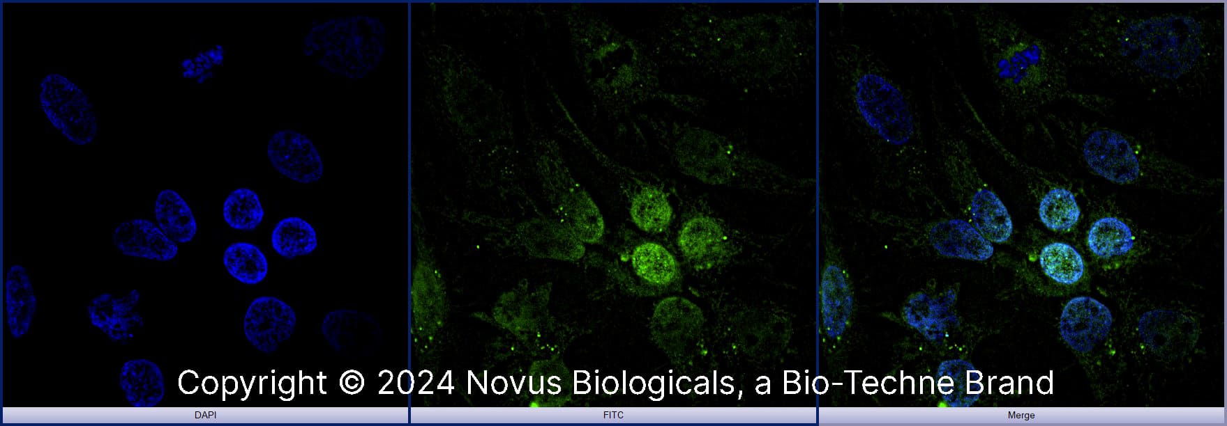
Scientific Data Images for Arginase 1/ARG1/liver Arginase Antibody - BSA Free
Western Blot: Arginase 1/ARG1/liver Arginase Antibody - BSA Free [NBP1-32731]
Western Blot: Arginase 1/ARG1/liver Arginase Antibody [NBP1-32731] - Non-transfected (-) and transfected (+) HepG2 whole cell extracts (30 ug) were separated by 10% SDS-PAGE, and the membrane was blotted with Arginase 1 antibody.Immunohistochemistry: Arginase 1/ARG1/liver Arginase Antibody - BSA Free [NBP1-32731]
Immunohistochemistry: Arginase 1/ARG1/liver Arginase Antibody [NBP1-32731] - TRP53-regulated EndMT modulates the M1 and M2 populations of increased TAMs after radiotherapy. Immunofluorescence detection of F4/80, CD31, and Arg1, in KP tumours from WT and EC-p53KO mice, with or without irradiation (23 days after irradiation). Image collected and cropped by Citeab from the following publication (Tumour-vasculature development via endothelial-to-mesenchymal transition after radiotherapy controls CD44v6+ cancer cell and macrophage polarization. Nat Commun (2018) licensed under a CC-BY license.Simple Western: Arginase 1/ARG1/liver Arginase AntibodyBSA Free [NBP1-32731]
Simple Western: Arginase 1/ARG1/liver Arginase Antibody [NBP1-32731] - Simple Western lane view shows a specific band for ARG1 in human and mouse Liver lysate using ARG1 antibody (NBP1-32731) at 25 ug/ml. This experiment was performed under reducing conditions using the 12-230 kDa separation system.Applications for Arginase 1/ARG1/liver Arginase Antibody - BSA Free
Application
Recommended Usage
Flow Cytometry
5 - 10 ug/ml
Immunocytochemistry/ Immunofluorescence
1:10 - 1:500
Immunohistochemistry
1:100-1:1000
Immunohistochemistry-Frozen
1:10 - 1:500
Immunohistochemistry-Paraffin
1:100-1:1000
Immunoprecipitation
1:10 - 1:500
Western Blot
1:1000-1:50000
Reviewed Applications
Read 1 review rated 4 using NBP1-32731 in the following applications:
Formulation, Preparation, and Storage
Purification
Immunogen affinity purified
Formulation
PBS
Format
BSA Free
Preservative
0.02% Sodium Azide
Concentration
1.0 mg/ml
Shipping
The product is shipped with polar packs. Upon receipt, store it immediately at the temperature recommended below.
Stability & Storage
Aliquot and store at -20C or -80C. Avoid freeze-thaw cycles.
Background: Arginase 1/ARG1
Arginase and nitric oxidase synthase (NOS) compete for the same L-arginine substrate, creating a delicate balance between pathways (1). Furthermore, bioavailability of L-arginine and ARG1 expression has been implicated in several pathologies including vascular disease, neuronal disease, cardiovascular disease, immune dysfunction, inflammation, and cancer (1,3-5). For instance, ARG1 functions as a macrophage marker, defining the M2 population, while inducible nitric oxide synthase (iNOS) characterizes the M1 population; impaired M1/M2 polarization and changes in ARG1 expression is observed in diseases such as arteriogenesis, asthma, pulmonary fibrosis, and inflammatory bowel disease (1,3). In humans, arginase deficiency, known as argininemia, is an autosomal recessive metabolic disorder characterized by elevated ammonia (hyperammonemia) levels and arginine accumulation (6). Given that many arginase-associated diseases are characterized by upregulation in expression of ARG1, ARG2, or both, arginase inhibitors are currently being studied as a potential therapeutic approach (1,4).
References
1. S Clemente, G., van Waarde, A., F Antunes, I., Domling, A., & H Elsinga, P. (2020). Arginase as a Potential Biomarker of Disease Progression: A Molecular Imaging Perspective. International Journal of Molecular Sciences. https://doi.org/10.3390/ijms21155291
2. Uniprot (P05089)
3. Kieler, M., Hofmann, M., & Schabbauer, G. (2021). More than just protein building blocks: How amino acids and related metabolic pathways fuel macrophage polarization. The FEBS Journal. Advance online publication. https://doi.org/10.1111/febs.15715
4. Shosha, E., Fouda, A. Y., Narayanan, S. P., Caldwell, R. W., & Caldwell, R. B. (2020). Is the Arginase Pathway a Novel Therapeutic Avenue for Diabetic Retinopathy?. Journal of Clinical Medicine. https://doi.org/10.3390/jcm9020425
5. Correale J. (2021). Immunosuppressive Amino-Acid Catabolizing Enzymes in Multiple Sclerosis. Frontiers in Immunology. https://doi.org/10.3389/fimmu.2020.600428
6. Morales, J. A., & Sticco, K. L. (2020). Arginase Deficiency. In StatPearls. StatPearls Publishing.
Long Name
Liver-Type Arginase
Alternate Names
AI, ARG1, Arginase-1, Liver Arginase, PGIF, Type I Arginase
Entrez Gene IDs
383 (Human)
Gene Symbol
ARG1
UniProt
Additional Arginase 1/ARG1 Products
Product Documents for Arginase 1/ARG1/liver Arginase Antibody - BSA Free
Product Specific Notices for Arginase 1/ARG1/liver Arginase Antibody - BSA Free
This product is for research use only and is not approved for use in humans or in clinical diagnosis. Primary Antibodies are guaranteed for 1 year from date of receipt.
Loading...
Loading...
Loading...
Loading...
Loading...
Loading...
![Immunohistochemistry: Arginase 1/ARG1/liver Arginase Antibody - BSA Free [NBP1-32731] Immunohistochemistry: Arginase 1/ARG1/liver Arginase Antibody - BSA Free [NBP1-32731]](https://resources.bio-techne.com/images/products/Arginase-1-ARG1-liver-Arginase-Antibody-Immunohistochemistry-NBP1-32731-img0033.jpg)
![Simple Western: Arginase 1/ARG1/liver Arginase AntibodyBSA Free [NBP1-32731] Simple Western: Arginase 1/ARG1/liver Arginase AntibodyBSA Free [NBP1-32731]](https://resources.bio-techne.com/images/products/Arginase-1-ARG1-liver-Arginase-Antibody-Simple-Western-NBP1-32731-img0029.jpg)
![Flow Cytometry: Arginase 1/ARG1/liver Arginase Antibody - BSA Free [NBP1-32731] Flow Cytometry: Arginase 1/ARG1/liver Arginase Antibody - BSA Free [NBP1-32731]](https://resources.bio-techne.com/images/products/Arginase-1-ARG1-liver-Arginase-Antibody-Flow-Cytometry-NBP1-32731-img0032.jpg)
![Western Blot: Arginase 1/ARG1/liver Arginase AntibodyBSA Free [NBP1-32731] Western Blot: Arginase 1/ARG1/liver Arginase AntibodyBSA Free [NBP1-32731]](https://resources.bio-techne.com/images/products/Arginase-1-ARG1-liver-Arginase-Antibody-Western-Blot-NBP1-32731-img0014.jpg)
![Immunoprecipitation: Arginase 1/ARG1/liver Arginase Antibody - BSA Free [NBP1-32731] Immunoprecipitation: Arginase 1/ARG1/liver Arginase Antibody - BSA Free [NBP1-32731]](https://resources.bio-techne.com/images/products/Arginase-1-ARG1-liver-Arginase-Antibody-Immunoprecipitation-NBP1-32731-img0015.jpg)
![Western Blot: Arginase 1/ARG1/liver Arginase AntibodyBSA Free [NBP1-32731] Western Blot: Arginase 1/ARG1/liver Arginase AntibodyBSA Free [NBP1-32731]](https://resources.bio-techne.com/images/products/Arginase-1-ARG1-liver-Arginase-Antibody-Western-Blot-NBP1-32731-img0020.jpg)
![Immunocytochemistry/ Immunofluorescence: Arginase 1/ARG1/liver Arginase Antibody - BSA Free [NBP1-32731] Immunocytochemistry/ Immunofluorescence: Arginase 1/ARG1/liver Arginase Antibody - BSA Free [NBP1-32731]](https://resources.bio-techne.com/images/products/Arginase-1-ARG1-liver-Arginase-Antibody-Immunocytochemistry-Immunofluorescence-NBP1-32731-img0034.jpg)
![Western Blot: Arginase 1/ARG1/liver Arginase AntibodyBSA Free [NBP1-32731] Western Blot: Arginase 1/ARG1/liver Arginase AntibodyBSA Free [NBP1-32731]](https://resources.bio-techne.com/images/products/Arginase-1-ARG1-liver-Arginase-Antibody-Western-Blot-NBP1-32731-img0018.jpg)
![Immunocytochemistry/ Immunofluorescence: Arginase 1/ARG1/liver Arginase Antibody - BSA Free [NBP1-32731] Immunocytochemistry/ Immunofluorescence: Arginase 1/ARG1/liver Arginase Antibody - BSA Free [NBP1-32731]](https://resources.bio-techne.com/images/products/Arginase-1-ARG1-liver-Arginase-Antibody-Immunocytochemistry-Immunofluorescence-NBP1-32731-img0009.jpg)
![Immunocytochemistry/ Immunofluorescence: Arginase 1/ARG1/liver Arginase Antibody - BSA Free [NBP1-32731] Immunocytochemistry/ Immunofluorescence: Arginase 1/ARG1/liver Arginase Antibody - BSA Free [NBP1-32731]](https://resources.bio-techne.com/images/products/Arginase-1-ARG1-liver-Arginase-Antibody-Immunocytochemistry-Immunofluorescence-NBP1-32731-img0027.jpg)
![Immunohistochemistry-Paraffin: Arginase 1/ARG1/liver Arginase Antibody - BSA Free [NBP1-32731] Immunohistochemistry-Paraffin: Arginase 1/ARG1/liver Arginase Antibody - BSA Free [NBP1-32731]](https://resources.bio-techne.com/images/products/Arginase-1-ARG1-liver-Arginase-Antibody-Immunohistochemistry-Paraffin-NBP1-32731-img0010.jpg)
![Immunohistochemistry-Paraffin: Arginase 1/ARG1/liver Arginase Antibody - BSA Free [NBP1-32731] Immunohistochemistry-Paraffin: Arginase 1/ARG1/liver Arginase Antibody - BSA Free [NBP1-32731]](https://resources.bio-techne.com/images/products/Arginase-1-ARG1-liver-Arginase-Antibody-Immunohistochemistry-Paraffin-NBP1-32731-img0026.jpg)
![Immunohistochemistry-Paraffin: Arginase 1/ARG1/liver Arginase Antibody - BSA Free [NBP1-32731] Immunohistochemistry-Paraffin: Arginase 1/ARG1/liver Arginase Antibody - BSA Free [NBP1-32731]](https://resources.bio-techne.com/images/products/Arginase-1-ARG1-liver-Arginase-Antibody-Immunohistochemistry-Paraffin-NBP1-32731-img0028.jpg)
![Flow Cytometry: Arginase 1/ARG1/liver Arginase Antibody - BSA Free [NBP1-32731] Flow Cytometry: Arginase 1/ARG1/liver Arginase Antibody - BSA Free [NBP1-32731]](https://resources.bio-techne.com/images/products/Arginase-1-ARG1-liver-Arginase-Antibody-Flow-Cytometry-NBP1-32731-img0025.jpg)
![Flow Cytometry: Arginase 1/ARG1/liver Arginase Antibody - BSA Free [NBP1-32731] Flow Cytometry: Arginase 1/ARG1/liver Arginase Antibody - BSA Free [NBP1-32731]](https://resources.bio-techne.com/images/products/Arginase-1-ARG1-liver-Arginase-Antibody-Flow-Cytometry-NBP1-32731-img0030.jpg)
![Western Blot: Arginase 1/ARG1/liver Arginase Antibody [NBP1-32731] - Arginase 1/ARG1/liver Arginase Antibody - BSA Free](https://resources.bio-techne.com/images/products/nbp1-32731_rabbit-polyclonal-arginase-1-arg1-liver-arginase-antibody-862023835350.jpg)
![Flow Cytometry: Arginase 1/ARG1/liver Arginase Antibody - BSA Free [NBP1-32731] - Arginase 1/ARG1/liver Arginase Antibody - BSA Free](https://resources.bio-techne.com/images/products/nbp1-32731_rabbit-polyclonal-arginase-1-arg1-liver-arginase-antibody-271220231253328.jpg)
![Immunocytochemistry/ Immunofluorescence: Arginase 1/ARG1/liver Arginase Antibody - BSA Free [NBP1-32731] - Arginase 1/ARG1/liver Arginase Antibody - BSA Free](https://resources.bio-techne.com/images/products/nbp1-32731_rabbit-polyclonal-arginase-1-arg1-liver-arginase-antibody-310202416165649.jpg)
