Bestrophin 1 Antibody (E6-6) - BSA Free
Novus Biologicals, part of Bio-Techne | Catalog # NB300-164

![Western Blot: Bestrophin 1 Antibody (E6-6)BSA Free [NB300-164] Western Blot: Bestrophin 1 Antibody (E6-6)BSA Free [NB300-164]](https://resources.bio-techne.com/images/products/Bestrophin-1-Antibody-E6-6-BSA-Free-Western-Blot-NB300-164-img0007.jpg)
Conjugate
Catalog #
Forumulation
Catalog #
Key Product Details
Validated by
Knockout/Knockdown
Species Reactivity
Validated:
Human, Mouse, Porcine, Canine, Primate, Rat (Negative)
Cited:
Human, Mouse, Rat, Porcine, Canine, Primate
Applications
Validated:
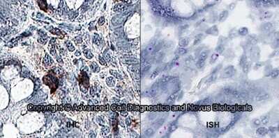
Dual RNAscope ISH-IHC, Immunocytochemistry/ Immunofluorescence, Immunohistochemistry, Immunohistochemistry-Frozen, Immunohistochemistry-Paraffin, Immunoprecipitation, Knockout Validated, Proximity Ligation Assay, Western Blot
Cited:
IF/ICC, IF/IHC, Immunocytochemistry/ Immunofluorescence, Immunohistochemistry-Frozen, Immunohistochemistry-Paraffin, Immunoprecipitation, Western Blot
Label
Unconjugated
Antibody Source
Monoclonal Mouse IgG1 kappa Clone # E6-6
Format
BSA Free
Concentration
1.0 mg/ml
Product Specifications
Immunogen
Synthetic peptide conjugated to KLH corresponding to the C-terminus of human Bestrophin 1 (KDHMDPYWALENRDEAHS)
Reactivity Notes
Human, Primate, Porcine reactivity reported in scientific literature (PMID: 11050159). Use in Mouse reported in scientific literature (PMID:32791386).
Clonality
Monoclonal
Host
Mouse
Isotype
IgG1 kappa
Scientific Data Images for Bestrophin 1 Antibody (E6-6) - BSA Free
Western Blot: Bestrophin 1 Antibody (E6-6)BSA Free [NB300-164]
Western Blot: Bestrophin 1 Antibody (E6-6) - BSA Free [NB300-164] - Western blot analysis of the normal and mutant human Best1 protein in transiently transfected MDCK cells. Best1 proteins are detectable as a 68 kDa band in all transfected cells, but not in non-transfected controls (MDCK lane). Actin bands are shown to indicate equal loading of cell lysates. Image collected and cropped by CiteAb from the following publication (https://www.mdpi.com/1422-0067/14/7/15121), licensed under a CC-BY license.Immunocytochemistry/ Immunofluorescence: Bestrophin 1 Antibody (E6-6) - BSA Free [NB300-164]
Immunocytochemistry/Immunofluorescence: Bestrophin 1 Antibody (E6-6) - BSA Free [NB300-164] - X-Z confocal single image scan of transiently transfected cells with different BEST1 cDNA constructs showing mislocalization of mutants Y85H, Q96R, L100R and Y227N. Cells were stained for Best1 (green), beta-catenin (red) and nuclei (blue). Scale bar = 10 um. Image collected and cropped by CiteAb from the following publication (https://www.mdpi.com/1422-0067/14/7/15121), licensed under a CC-BY license.Immunohistochemistry-Paraffin: Bestrophin 1 Antibody (E6-6) - BSA Free [NB300-164]
Immunohistochemistry-Paraffin: Bestrophin 1 Antibody (E6-6) - BSA Free [NB300-164] - Bestrophin 1 was detected in immersion fixed paraffin sections of human small intestine using t Mouse Anti-Human Bestrophin 1 Monoclonal Antibody (Catalog # NB300-164) at 5 ug/mL for 1 hour at room temperature followed by incubation with the Anti-Mouse IgG VisUCyte™ HRP Polymer Antibody (Catalog # VC001). Tissue was stained using DAB (brown) and counterstained with hematoxylin (blue). Specific staining was localized to the cell surface and extracellular.Applications for Bestrophin 1 Antibody (E6-6) - BSA Free
Application
Recommended Usage
Immunohistochemistry
reported in scientific literature (PMID 30048622)
Immunohistochemistry-Paraffin
reported in scientific literature (PMID 24345323)
Proximity Ligation Assay
reported in scientific literature (PMID 27519691)
Western Blot
1:1000
Application Notes
In Western blot, this antibody recognizes a band at ~68 kDa representing Bestrophin. Please see protocol for treatment of cell extracts. The observed molecular weight of the protein may vary from the listed predicted molecular weight due to post translational modifications, post translation cleavages, relative charges, and other experimental factors.
Reviewed Applications
Read 1 review rated 5 using NB300-164 in the following applications:
Formulation, Preparation, and Storage
Purification
Protein A or G purified
Formulation
PBS
Format
BSA Free
Preservative
0.02% Sodium Azide
Concentration
1.0 mg/ml
Shipping
The product is shipped with polar packs. Upon receipt, store it immediately at the temperature recommended below.
Stability & Storage
Aliquot and store at -20C or -80C. Avoid freeze-thaw cycles.
Background: Bestrophin 1
Alternate Names
ARB, BEST, Best disease, bestrophin 1, bestrophin-1, BMD, TU15B, vitelliform macular dystrophy 2, Vitelliform macular dystrophy protein 2, VMD2RP50
Gene Symbol
BEST1
UniProt
Additional Bestrophin 1 Products
Product Documents for Bestrophin 1 Antibody (E6-6) - BSA Free
Product Specific Notices for Bestrophin 1 Antibody (E6-6) - BSA Free
This product is for research use only and is not approved for use in humans or in clinical diagnosis. Primary Antibodies are guaranteed for 1 year from date of receipt.
Loading...
Loading...
Loading...
Loading...
Loading...
![Immunocytochemistry/ Immunofluorescence: Bestrophin 1 Antibody (E6-6) - BSA Free [NB300-164] Immunocytochemistry/ Immunofluorescence: Bestrophin 1 Antibody (E6-6) - BSA Free [NB300-164]](https://resources.bio-techne.com/images/products/Bestrophin-1-Antibody-E6-6-BSA-Free-Immunocytochemistry-Immunofluorescence-NB300-164-img0008.jpg)
![Immunohistochemistry-Paraffin: Bestrophin 1 Antibody (E6-6) - BSA Free [NB300-164] Immunohistochemistry-Paraffin: Bestrophin 1 Antibody (E6-6) - BSA Free [NB300-164]](https://resources.bio-techne.com/images/products/Bestrophin-1-Antibody-E6-6-BSA-Free-Immunohistochemistry-Paraffin-NB300-164-img0005.jpg)
![Western Blot: Bestrophin 1 Antibody (E6-6)BSA Free [NB300-164] Western Blot: Bestrophin 1 Antibody (E6-6)BSA Free [NB300-164]](https://resources.bio-techne.com/images/products/Bestrophin-1-Antibody-E6-6-BSA-Free-Western-Blot-NB300-164-img0003.jpg)
![Immunohistochemistry-Paraffin: Bestrophin 1 Antibody (E6-6) - BSA Free [NB300-164] Immunohistochemistry-Paraffin: Bestrophin 1 Antibody (E6-6) - BSA Free [NB300-164]](https://resources.bio-techne.com/images/products/Bestrophin-1-Antibody-E6-6-BSA-Free-Immunohistochemistry-Paraffin-NB300-164-img0004.jpg)
![Western Blot: Bestrophin 1 Antibody (E6-6) - BSA Free [NB300-164] Knockout Validated: Bestrophin 1 Antibody (E6-6) - BSA Free [NB300-164]](https://resources.bio-techne.com/images/products/Bestrophin-1-Antibody-E6-6-BSA-Free-Knockout-Validated-NB300-164-img0009.jpg)

![Western Blot: Bestrophin 1 Antibody (E6-6) - BSA Free [NB300-164] - Bestrophin 1 Antibody (E6-6) - BSA Free](https://resources.bio-techne.com/images/products/nb300-164_mouse-monoclonal-bestrophin-1-antibody-e6-6-21020242346812.jpg)
![Western Blot: Bestrophin 1 Antibody (E6-6) - BSA Free [NB300-164] - Bestrophin 1 Antibody (E6-6) - BSA Free](https://resources.bio-techne.com/images/products/nb300-164_mouse-monoclonal-bestrophin-1-antibody-e6-6-31020241534336.jpg)
![Immunocytochemistry/ Immunofluorescence: Bestrophin 1 Antibody (E6-6) - BSA Free [NB300-164] - Bestrophin 1 Antibody (E6-6) - BSA Free](https://resources.bio-techne.com/images/products/nb300-164_mouse-monoclonal-bestrophin-1-antibody-e6-6-310202415363746.jpg)
![Western Blot: Bestrophin 1 Antibody (E6-6) - BSA Free [NB300-164] - Bestrophin 1 Antibody (E6-6) - BSA Free](https://resources.bio-techne.com/images/products/nb300-164_mouse-monoclonal-bestrophin-1-antibody-e6-6-310202415284544.jpg)
![Immunocytochemistry/ Immunofluorescence: Bestrophin 1 Antibody (E6-6) - BSA Free [NB300-164] - Bestrophin 1 Antibody (E6-6) - BSA Free](https://resources.bio-techne.com/images/products/nb300-164_mouse-monoclonal-bestrophin-1-antibody-e6-6-31020241535684.jpg)
![Western Blot: Bestrophin 1 Antibody (E6-6) - BSA Free [NB300-164] - Bestrophin 1 Antibody (E6-6) - BSA Free](https://resources.bio-techne.com/images/products/nb300-164_mouse-monoclonal-bestrophin-1-antibody-e6-6-31020241537190.jpg)
![Western Blot: Bestrophin 1 Antibody (E6-6) - BSA Free [NB300-164] - Bestrophin 1 Antibody (E6-6) - BSA Free](https://resources.bio-techne.com/images/products/nb300-164_mouse-monoclonal-bestrophin-1-antibody-e6-6-31020241539730.jpg)
![Western Blot: Bestrophin 1 Antibody (E6-6) - BSA Free [NB300-164] - Bestrophin 1 Antibody (E6-6) - BSA Free](https://resources.bio-techne.com/images/products/nb300-164_mouse-monoclonal-bestrophin-1-antibody-e6-6-31020241535628.jpg)
![Immunocytochemistry/ Immunofluorescence: Bestrophin 1 Antibody (E6-6) - BSA Free [NB300-164] - Bestrophin 1 Antibody (E6-6) - BSA Free](https://resources.bio-techne.com/images/products/nb300-164_mouse-monoclonal-bestrophin-1-antibody-e6-6-310202415395959.jpg)
![Immunocytochemistry/ Immunofluorescence: Bestrophin 1 Antibody (E6-6) - BSA Free [NB300-164] - Bestrophin 1 Antibody (E6-6) - BSA Free](https://resources.bio-techne.com/images/products/nb300-164_mouse-monoclonal-bestrophin-1-antibody-e6-6-310202415175245.jpg)
![Immunocytochemistry/ Immunofluorescence: Bestrophin 1 Antibody (E6-6) - BSA Free [NB300-164] - Bestrophin 1 Antibody (E6-6) - BSA Free](https://resources.bio-techne.com/images/products/nb300-164_mouse-monoclonal-bestrophin-1-antibody-e6-6-31020241621835.jpg)