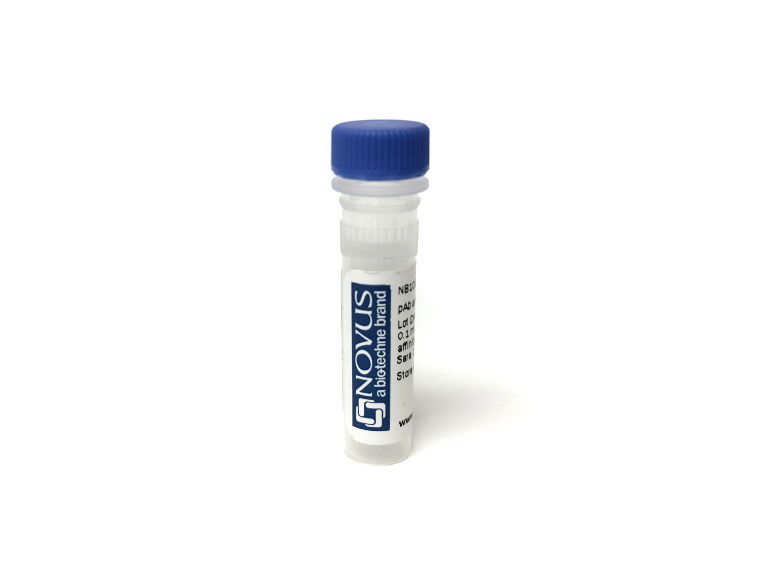
c-Myc Antibody (9E10.3) [CoraFluor™ 1]
Novus Biologicals, part of Bio-Techne | Catalog # NBP2-47738CL1


Conjugate
Catalog #
Forumulation
Catalog #
Key Product Details
Species Reactivity
Human, Chicken (Negative), Mouse (Negative), Rat (Negative)
Applications
CyTOF-ready, Flow Cytometry, Immunocytochemistry/ Immunofluorescence, Immunofluorescence, Immunohistochemistry, Immunohistochemistry-Paraffin
Label
CoraFluor 1
Antibody Source
Monoclonal Mouse IgG1 kappa Clone # 9E10.3
Concentration
Please see the vial label for concentration. If unlisted please contact technical services.
Product Specifications
Immunogen
A synthetic peptide, corresponding to aa 408-439 (AEEQKLISEEDLLRKRREQLKHKLEQLRNSCA) from C-terminus of human c-Myc, coupled to KLH. (Uniprot: P01106)
Epitope
aa 410-419 (EQKLISEEDL)
Reactivity Notes
Does not react with Mouse, Rat or Chicken.
Localization
Nuclear
Specificity
It recognizes a transcription factor of 64-67kDa, identified as c-myc. Its epitope spans between aa 410-419 (EQKLISEEDL) which is a specific portion of an alpha helical region of human c-myc protein. This monoclonal antibody shows no cross-reaction with v-myc. c-myc is involved in the control of cell proliferation and differentiation and is amplified and/or overexpressed in a variety of tumors. Over-expression of c-myc protein occurs frequently in luminal cells of prostate intraepithelial neoplasia as well as in most primary carcinomas and metastatic disease.
Clonality
Monoclonal
Host
Mouse
Isotype
IgG1 kappa
Description
CoraFluor(TM) 1 is a high performance terbium-based TR-FRET (Time-Resolved Fluorescence Resonance Energy Transfer) or TRF (Time-Resolved Fluorescence) donor for high throughput assay development. CoraFluor(IM) 1 absorbs UV light at approximately 340 nm, and emits at approximately 490 nm, 545 nm, 585 nm and 620 nm. It is compatible with common acceptor dyes that absorb at the emission wavelengths of CoraFluor(TM) 1. CoraFluor(TM) 1 can be used for the development of robust and scalable TR-FRET binding assays such as target engagement, ternary complex, protein-protein interaction and protein quantification assays.
Applications for c-Myc Antibody (9E10.3) [CoraFluor™ 1]
Application
Recommended Usage
CyTOF-ready
Optimal dilutions of this antibody should be experimentally determined.
Flow Cytometry
Optimal dilutions of this antibody should be experimentally determined.
Immunocytochemistry/ Immunofluorescence
Optimal dilutions of this antibody should be experimentally determined.
Immunofluorescence
Optimal dilutions of this antibody should be experimentally determined.
Immunohistochemistry
Optimal dilutions of this antibody should be experimentally determined.
Immunohistochemistry-Paraffin
Optimal dilutions of this antibody should be experimentally determined.
Application Notes
Optimal dilution of this antibody should be experimentally determined.
Formulation, Preparation, and Storage
Purification
Protein A or G purified
Formulation
PBS
Preservative
No Preservative
Concentration
Please see the vial label for concentration. If unlisted please contact technical services.
Shipping
The product is shipped with polar packs. Upon receipt, store it immediately at the temperature recommended below.
Stability & Storage
Store at 4C in the dark. Do not freeze.
Background: c-Myc
A basic Helix-Loop-Helix, Leucine Zipper domain (bHLH/LZ), designated Max, specifically associates with c-Myc, N-Myc and L-Myc proteins. The Myc-Max complex binds to DNA in a sequence-specific manner under conditions where neither Max nor Myc exhibit appreciable binding. Max can also form heterodimers with other bHLH-Zip proteins, Mad and Mxi1. c-Myc plays a role in cell cycle progression, apoptosis, cellular transformation and angiogenesis (2). Mutations, overexpression, rearrangement and translocation of this gene have been associated with a variety of cancers including B-cell Lymphomas, acute myeloid leukemia, glioblastoma, stomach adenocarcinoma, and prostate adenocarcinoma (3).
References
1. Wilkinson, D. S., Tsai, W. W., Schumacher, M. A., & Barton, M. C. (2008). Chromatin-bound p53 anchors activated Smads and the mSin3A corepressor to confer transforming-growth-factor-beta-mediated transcription repression. Mol Cell Biol, 28(6), 1988-1998. doi:10.1128/mcb.01442-07
2. Pedrosa, A. R., Bodrug, N., Gomez-Escudero, J., Carter, E. P., Reynolds, L. E., Georgiou, P. N., . . . Hodivala-Dilke, K. M. (2019). Tumor Angiogenesis Is Differentially Regulated by Phosphorylation of Endothelial Cell Focal Adhesion Kinase Tyrosines-397 and -861. Cancer Res, 79(17), 4371-4386. doi:10.1158/0008-5472.Can-18-3934
3. Nagasaka, M., Tsuzuki, K., Ozeki, Y., Tokugawa, M., Ohoka, N., Inoue, Y., & Hayashi, H. (2019). Lysine-Specific Demethylase 1 (LSD1/KDM1A) Is a Novel Target Gene of c-Myc. Biol Pharm Bull, 42(3), 481-488. doi:10.1248/bpb.b18-00892
Long Name
v-Myc Avian Myelocytomatosis Viral Oncogene Homolog (Avian)
Alternate Names
cMyc, Myc, Myc2, Niard, Nird
Gene Symbol
MYC
Additional c-Myc Products
Product Documents for c-Myc Antibody (9E10.3) [CoraFluor™ 1]
Product Specific Notices for c-Myc Antibody (9E10.3) [CoraFluor™ 1]
CoraFluor (TM) is a trademark of Bio-Techne Corp. Sold for research purposes only under agreement from Massachusetts General Hospital. US patent 2022/0025254
This product is for research use only and is not approved for use in humans or in clinical diagnosis. Primary Antibodies are guaranteed for 1 year from date of receipt.
Loading...
Loading...
Loading...
Loading...
Loading...
Loading...