Canine CCL2/JE/MCP-1 Biotinylated Antibody
R&D Systems, part of Bio-Techne | Catalog # BAF1774


Key Product Details
Species Reactivity
Applications
Label
Antibody Source
Product Specifications
Immunogen
Gln24-Pro101
Accession # P52203
Specificity
Clonality
Host
Isotype
Scientific Data Images for Canine CCL2/JE/MCP-1 Biotinylated Antibody
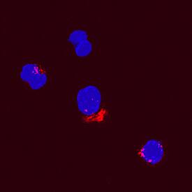
CCL2/JE/MCP‑1 in Canine PBMCs.
CCL2/JE/MCP-1 was detected in immersion fixed canine peripheral blood mononuclear cells (PBMCs) using Goat Anti-Canine CCL2/JE/MCP-1 Biotinylated Antigen Affinity-purified Polyclonal Antibody (Catalog # BAF1774) at 15 µg/mL for 3 hours at room temperature. Cells were stained using the NorthernLights™ 557-conjugated Streptavidin (red; Catalog # NL999) and counterstained with DAPI (blue). Specific staining was localized to cytoplasmic. View our protocol for Fluorescent ICC Staining of Non-adherent Cells.Applications for Canine CCL2/JE/MCP-1 Biotinylated Antibody
Immunocytochemistry
Sample: Immersion fixed canine peripheral blood mononuclear cells (PBMCs)
Western Blot
Sample: Recombinant Canine CCL2/JE/MCP-1 (Catalog # 1774-MC)
Canine CCL2/JE/MCP-1 Sandwich Immunoassay
Formulation, Preparation, and Storage
Purification
Reconstitution
Formulation
Shipping
Stability & Storage
- 12 months from date of receipt, -20 to -70 °C as supplied.
- 1 month, 2 to 8 °C under sterile conditions after reconstitution.
- 6 months, -20 to -70 °C under sterile conditions after reconstitution.
Background: CCL2/JE/MCP-1
Canine MCP-1 (monocyte chemotactic protein-1) is an 8 kDa member of the CC chemokine family of chemotactic factors (1, 2). It is synthesized as a 101 amino acid (aa) precursor that contains a 23 aa signal sequence and a 78 aa mature segment (3). It contains no potential N-linked glycosylation sites and is not known for any posttranslational modifications. Based on human studies, MCP-1 will primarily circulate as a monomer. Noncovalent dimers are likely to be found, however. MCP‑1 activity has been localized to the N-terminus (1). Cell types known to secrete MCP-1 are considerable in number, and include keratinocytes, fibroblasts, endothelium, osteoblasts, macrophages, mast cells, smooth muscle cells and astrocytes (1, 2). In the mature MCP-1 segment, there is 82% and 83% aa identity, canine to human and porcine MCP-1, respectively. When mature canine MCP-1 is compared to (125 aa) extended rodent MCP-1, there is 55% and 56% aa identity, canine to mouse and rat MCP-1, respectively. MCP-1 has three possible receptors. The first two are CCR2 (1) and CCR11 (4). The third receptor has only been identified in mice and is called L-CCR (5). Its function is unknown. MCP-1 is best known as a chemotactic agent for mononuclear cells. It also, however, induces enzyme and cytokine release in monocytes, NK cells, and lymphocytes and histamine release by basophils (1). Additionally, it is believed to reduce IL‑12 production by dendritic cells and promote a Th2 phenotype in CD4+ T cells (6).
References
- Coillie, E.V. et al. (1999) Cytokine Growth Factor Rev. 10:61.
- Yoshie, O. et al. (2001) Adv. Immunol. 78:57.
- Kumar, A.G. et al. (1997) Circulation 95:693.
- Biber, K. et al. (2003) J. Leukoc. Biol. 74:243.
- Luther, S.A. and J.G. Cyster (2001) Nat. Immunol. 2:102.
Alternate Names
Gene Symbol
UniProt
Additional CCL2/JE/MCP-1 Products
Product Documents for Canine CCL2/JE/MCP-1 Biotinylated Antibody
Product Specific Notices for Canine CCL2/JE/MCP-1 Biotinylated Antibody
For research use only