Canine/Equine CD44 Antibody
R&D Systems, part of Bio-Techne | Catalog # MAB5449


Key Product Details
Species Reactivity
Validated:
Cited:
Applications
Validated:
Cited:
Label
Antibody Source
Product Specifications
Immunogen
Accession # Q28284
Specificity
Clonality
Host
Isotype
Scientific Data Images for Canine/Equine CD44 Antibody
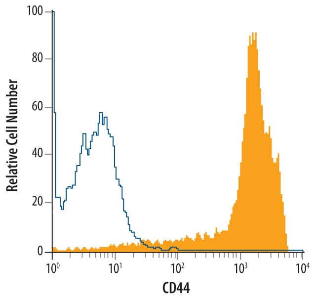
Detection of CD44 in Canine Monocytes by Flow Cytometry.
Canine blood-derived monocytes were stained with Mouse Anti-Canine/Equine CD44 Monoclonal Antibody (Catalog # MAB5449, filled histogram) or isotype control antibody (Catalog # MAB002, open histogram), followed by Allophycocyanin-conjugated Anti-Mouse IgG F(ab')2Secondary Antibody (Catalog # F0101B).Detection of CD44 in Equine PBMCs by Flow Cytometry.
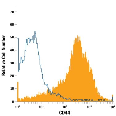
Equine peripheral blood mononuclear cells (PBMCs) were stained with Mouse Anti-Canine/Equine CD44 Monoclonal Antibody (Catalog # MAB5449, filled histogram) or isotype control antibody (Catalog # MAB002, open histogram), followed by Phycoerythrin-conjugated Anti-Mouse IgG Secondary Antibody (Catalog # F0102B).Detection of Equine CD44 by Flow Cytometry
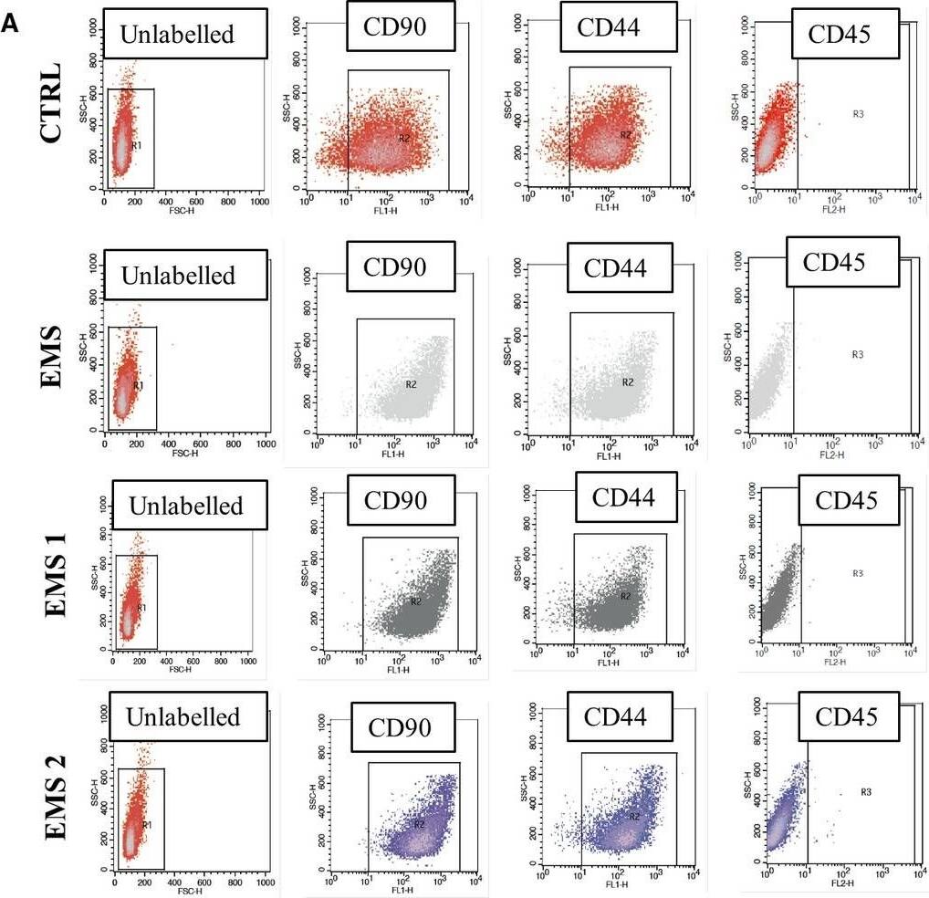
Immunophenotyping and multipotency assay. Representative dot plots from flow cytometry analysis (A). Expression of surface antigens were investigated in cells cultured in control and AZA/RES supplemented medium. Isolated cells were characterized by the expression of CD90 and CD44 while lacked the expression of CD45 hematopoietic marker (B). Multipotency of isolated cells was confirmed by three lineage differentiation (C). In order to confirm formation of proteoglycans during chondrogenesis cells were stained with Safranin O. Intracellular lipid droplets were visualized by Oil Red O while mineralized matrix formed during osteogenesis with Alizarin Red. Magnification ×100, scale bars: 250 μm. Results expressed as mean ± SD. Statistical significance indicated as asterisk (*) when comparing the result to ASCCTRL, and as hashtag (#) when comparing to ASCEMS. ###P < 0.001 Image collected and cropped by CiteAb from the following open publication (https://pubmed.ncbi.nlm.nih.gov/30370650), licensed under a CC-BY license. Not internally tested by R&D Systems.Applications for Canine/Equine CD44 Antibody
CyTOF-ready
Flow Cytometry
Sample: Canine blood‑derived monocytes and equine peripheral blood mononuclear cells (PBMCs)
Formulation, Preparation, and Storage
Purification
Reconstitution
Formulation
Shipping
Stability & Storage
- 12 months from date of receipt, -20 to -70 °C as supplied.
- 1 month, 2 to 8 °C under sterile conditions after reconstitution.
- 6 months, -20 to -70 °C under sterile conditions after reconstitution.
Background: CD44
Canine CD44 is a ubiquitously expressed 85‑90 kDa transmembrane glycoprotein that binds to hyaluronan and is involved in matrix adhesion, lymphocyte activation, and lymph node homing. The CD44 protein is expressed as a family of molecular isoforms generated by alternate splicing and variable posttranslational modification. Within the N‑terminal invariant portion of the ECD (aa 14‑191), canine CD44 shares 90%, 83%, and 82% identity with human, mouse, and rat CD44, respectively.
Alternate Names
Gene Symbol
UniProt
Additional CD44 Products
Product Documents for Canine/Equine CD44 Antibody
Product Specific Notices for Canine/Equine CD44 Antibody
For research use only

