Immunohistological Staining of Carbonic Anhydrase IX/CA9 in Endometrial Cancer
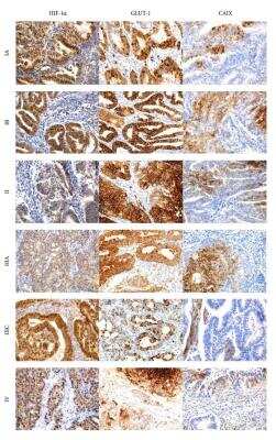
Immunohistochemical representative microphotographs representing the HIF-1alpha, GLUT-1, and CAIX expression in endometrial cancer according to FIGO classification (IA, IB, II, IIIA, IIIC, and IV). Primary objective magnification 20x. Image collected and cropped by CiteAb from the following publication (https://www.hindawi.com/journals/bmri/2014/616850/) licensed under a CC-BY license.
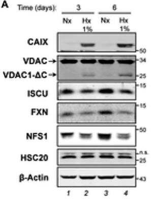
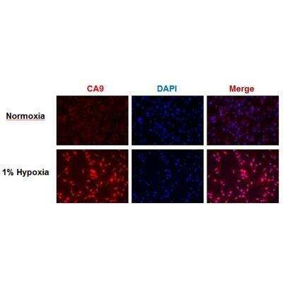
Western Blot Detection of Carbonic Anhydrase IX/CA9 in Hypoxic and Normoxic HeLa Cells
Hypoxias impact on proteins of the mitochondrial ISC assembly machinery. (A) Immunoblotting analyzed the total protein extracts from HeLa cells grown in normoxia (Nx, 21% O2) or hypoxia (Hx, 1% O2) conditions using VDACs poly antibody and anti-CAIX, -ISCU, -FXN, -NFS1, -HSC20 antibodies. Beta-Actin was used as loading control. Citation: Ferecatu I, Canal F, Fabbri L, Mazure NM, Bouton C, Golinelli-Cohen M-P (2018) Dysfunction in the mitochondrial Fe-S assembly machinery leads to formation of the chemoresistant truncated VDAC1 isoform without HIF-1 alpha activation. PLoS ONE 13(3): e0194782.
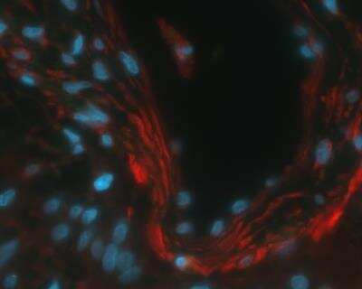
Immunofluorescent Staining of Carbonic Anhydrase IX/CA9 in Human Glioma U87 Cells
Analysis using the DyLight 488 conjugate of NB100-417. Staining of Carbonic Anhydrase IX (red) in human glioma U87 cells. DAPI counterstains nuclei (blue). ICC/IF image submitted by a verified customer review.
Detection of Carbonic Anhydrase IX/CA9 in HeLa Cell Lysate by Simple Western
Simple Western lane view shows a specific band for CAIX in 0.1 mg/mL of HeLa lysate. This experiment was performed under reducing conditions using the 12-230 kDa separation system.
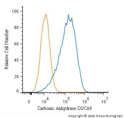
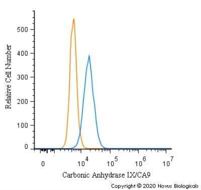
Flow Cytometry of U-87 MG Cells Stained with Carbonic Anhydrase IX/CA9 Antibody
An intracellular stain was performed on U-87 MG Cells with NB100-417 and a matched isotype control. Cells were fixed with 4% PFA and then permeablized with 0.1% saponin. Cells were incubated in an antibody dilution of 2.5 ug/mL for 30 minutes at room temperature, followed by Rabbit IgG APC-conjugated Secondary Antibody, (R&D Systems, F0111).
Western Blot Detection of Carbonic Anhydrase IX/CA9 in Human Breast Cancer MCF7 Cells
Western Blot analysis of Carbonic Anhydrase IX/CA9 Antibody on human breast cancer MCF7 cells. Image from verified customer review.
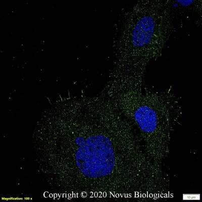
Immunocytochemistry/Immunofluorescence Staining of Carbonic Anhydrase IX/CA9 in A431 Cells
A431 cells were fixed in 4% paraformaldehyde for 10 minutes and permeabilized in 0.05% Triton X-100 in PBS for 5 minutes. The cells were incubated with anti-Carbonic Anhydrase IX/CA9 Antibody NB100-417 at 2 ug/ml overnight at 4C and detected with an anti-rabbit Dylight 488 (Green) at a 1:1000 dilution for 60 minutes. Nuclei were counterstained with DAPI (Blue). Cells were imaged using a 100X objective and digitally deconvolved.
Flow Cytometry of A431 Cells Stained with Carbonic Anhydrase IX/CA9 Antibody
An intracellular stain was performed on A431 cells with Carbonic Anhydrase IX/CA9 Antibody NB100-417 (blue) and a matched isotype control (orange). Cells were fixed with 4% PFA and then permeabilized with 0.1% saponin. Cells were incubated in an antibody dilution of 1.0 ug/mL for 30 minutes at room temperature, followed by Rabbit IgG (H+L) Cross-Adsorbed Secondary Antibody, Dylight 550 (SA5-10033, Thermo Fisher).
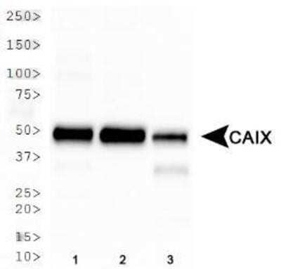
Western Blot Analysis of Carbonic Anhydrase IX/CA9 in Multiple Whole Cell Lysates
Analysis in 1) HeLa, 2) MDA-MB-231, and 3) A549 whole cell lysates. Specific bands were detected for Carbonic Anhydrase IX/CA9 at a molecular weight of 50 kDa.
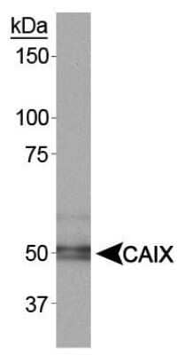
Detection of Carbonic Anhydrase IX/CA9 in Rat Renal Cortex by Western Blot
Analysis on rat renal cortex. A specific band was detected at a molecular weight of approximately 50 kDa.
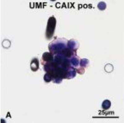
Staining of Carbonic Anhydrase IX/CA9 in CNHCs
Immunocytochemical analysis of CNHCs with antibodies against the RCC marker CAIX. Clusters of CNHCs cytomorphologically classified as uncertain malignant (-UMF) with cytoplasmic positive staining with antibodies against the RCC marker CAIX. Image collected and cropped by CiteAb from the following publication (https://translational-medicine.biomedcentral.com/articles/10.1186/1479-5876-11-214) licensed under a CC-BY license.
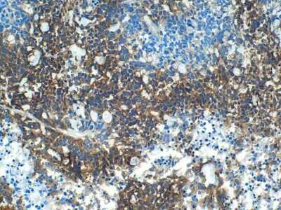
Immunohistochemical Staining of Carbonic Anhydrase IX/CA9 in Frozen Mouse Xenografted Human Colon Carcinoma
Analysis of human colon carcinoma, xenografted in mice. IHC-Fr image submitted by a verified customer review.
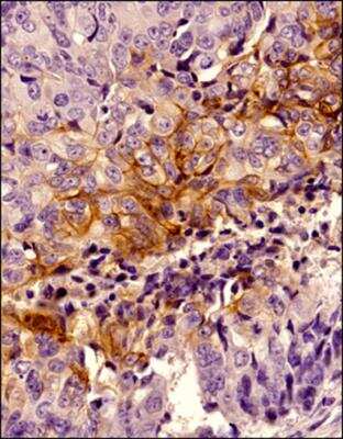
Immunohistochemical Staining of Carbonic Anhydrase IX/CA9 in Paraffin Embedded Human Breast Cancer
IHC analysis of a FFPE tissue section of human breast cancer using CAIX antibody at 1:1000 dilution. The primary antibody bound to CAIX antigens in the tissue section was detected using a HRP labeled secondary antibody and DAB reagent. Nuclei of the cells were counterstained with hematoxylin. This CAIX antibody generated an expected cytoplasmic staining of CAIX protein with an intense signal around the cellular membranes in tumor cores. The latter are more likely to be hypoxic in growing tumors which signifies that the observed CAIX staining is specific.
Immunohistochemical Staining of Carbonic Anhydrase IX/CA9 in Paraffin Embedded Human Stomach Tissue
Strong cytoplasmic and membranous CA9 staining in glandular cells of human stomach tissue. Citrate buffer pH 6 antigen retrieval. Antibody at 1:250 dilution and detected with an Alexa Fluor 546 labeled goat-anti-rabbit secondary antibody. Nuclei detected with DAPI. IHC-P image submitted by a verified customer review.
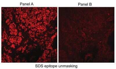
Immunohistological Detection of Carbonic Anhydrase IX/CA9 in Human RCC Tumor Cryosections
Immunofluorescence of human RCC tumor cryosections using NB100-417 (Panel A). Panel B shows staining with normal rabbit serum.
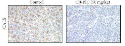
Immunohistochemical Staining of Carbonic Anhydrase IX/CA9 in Control and CB-PIC Treated Colorectal Cancer
Colorectal cancer xenograft growth was suppressed by CB-PIC(20 and 50 mg/kg body weight) in female athymic nude mice. Starting three days after SW620 cell inoculation, CB-PIC (20 and 50 mg/kg body weight) was injected in abdomen with 4% Tween 20 as vehicle once daily. Representative examples of immunohistochemical staining for CA IX in tumor section. Image collected and cropped by CiteAb from the following publication (https://www.hindawi.com/journals/ecam/2013/974313/), licensed under a CC-BY license.
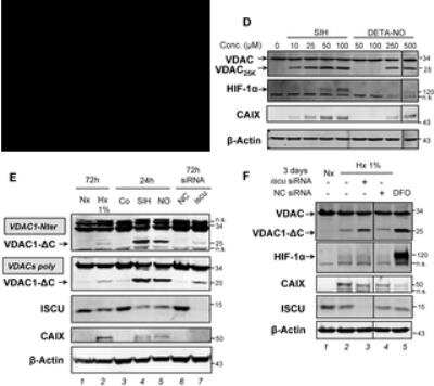
Western Blot Detection of Carbonic Anhydrase IX/CA9 in HeLa Cells Under Multiple Conditions and Treatments
Iron depletion and nitric oxide stress and Iron depletion induce the truncated VDAC1 form accumulation. (D) Total protein extracts were analyzed by immunoblotting using VDACs poly antibody and anti-HIF-1 alpha and -CAIX antibodies. (E) Immunoblotting was used to analyze total protein extracts using antibodies against the three VDAC isoforms. (F) HeLa cells were grown in hypoxia (Hx, 1% O2) conditions and transfected with iscu- or NC-siRNA for 3 days, or grown in normoxia (Nx, 21% O2), some treated with DFO for 16 h. Western Blots analyzed total proteins using VDACs poly antibody and anti-HIF-1 alpha, -CAIX, -ISCU antibodies. Citation: Ferecatu I, Canal F, Fabbri L, Mazure NM, Bouton C, Golinelli-Cohen M-P (2018) Dysfunction in the mitochondrial Fe-S assembly machinery leads to formation of the chemoresistant truncated VDAC1 isoform without HIF-1 alpha activation. PLoS ONE 13(3): e0194782.
Immunohistochemistry: Carbonic Anhydrase IX/CA9 Antibody - BSA Free [NB100-417] -
Immunohistochemical representative microphotographs representing the HIF-1 alpha, GLUT-1, and CAIX expression in endometrial cancer according to FIGO classification (IA, IB, II, IIIA, IIIC, and IV). Primary objective magnification 20x.
Immunohistochemistry: Carbonic Anhydrase IX/CA9 Antibody - BSA Free [NB100-417] -
The images are representative of immunohistochemistry staining of nuclear staining of proliferation marker Ki67, membrane protein CD 44, and CA IX. The images of scaffold culture (left panel) are compared with cell pellet paraffin section (right panel). The scaffold dissolved in xylene presented as a clear region marked ∗. All scale bars are 100 μm, magnification 40x.
Immunohistochemistry: Carbonic Anhydrase IX/CA9 Antibody - BSA Free [NB100-417] -
Chemosensitivity of the CTC SCLC lines (IC50, mean ± SD) and immunohistochemistry of sections of tumorospheres. All differences between single cells (SC) and tumorospheres (TS) are statistically significant. Immunohistochemical staining of sections of UHGc5, BHGc16 and BHGc26 CTCs was performed using antibodies directed to CD56, CHGA, CAIX and Ki67, respectively.
Western Blot: Carbonic Anhydrase IX/CA9 Antibody - BSA Free [NB100-417] -
Western Blot: Carbonic Anhydrase IX/CA9 Antibody - BSA Free [NB100-417] - Analysis of protein levels of genes with increased transcription in hypoxia, IOX2 & VH032.HIF targets were increased in hypoxia, VH298 & FG-4592. 0.05% DMSO (vehicle control), 1% O2 (hypoxia), 100 µM VH298 & 50 µM FG-4592 were introduced to (A) HeLa or HFF for 24 hours & (B) HeLa for indicated time. Protein levels were analysed by immunoblotting using antibodies against indicated proteins, with beta-Actin as loading control. The blots shown are representative of three independent experiments. * indicates longer exposure. Image collected & cropped by CiteAb from the following publication (https://pubmed.ncbi.nlm.nih.gov/30801039), licensed under a CC-BY license. Not internally tested by Novus Biologicals.
Immunohistochemistry-Paraffin: Carbonic Anhydrase IX/CA9 Antibody - BSA Free [NB100-417] -
Immunohistochemistry-Paraffin: Carbonic Anhydrase IX/CA9 Antibody - BSA Free [NB100-417] - IGF1R & CA9 expression in PDAC.(A) Representative staining of CA9 cells quantified on a scoring system of 0–3 according to staining intensity (original magnification ×200). CA9 was mainly expressed in the cell membrane of PDAC cells. (B) IGF1R was mainly expressed in the cytoplasm of PDAC cells. (C) Association between IGF1Rexpression & CA9 expression in 120 pancreas cancer. A significant correlation was found between IGF1R expression level was significantly correlated with CA9 expression in PDAC cells (p < 0.01, rs = 0.649; Spearman’s rank sum test). The size of circle indicates the number of PDAC patients. Image collected & cropped by CiteAb from the following publication (https://pubmed.ncbi.nlm.nih.gov/27487118), licensed under a CC-BY license. Not internally tested by Novus Biologicals.
Immunohistochemistry: Carbonic Anhydrase IX/CA9 Antibody - BSA Free [NB100-417] -
Immunohistochemistry: Carbonic Anhydrase IX/CA9 Antibody - BSA Free [NB100-417] - Representative human colorectal cancer specimen. Immunohistochemistry of CA9 & GGT1 in mLNs. In these cases, both CA9 & GGT1 were expressed, suggesting that GGT1 expression is related to hypoxia in mLNs. Scale bar, 500 μm. Black arrowheads: GGT of cancer cells, white arrowheads: GGT surrounding cancer cells, arrow: accumulation of GGT. mLNs: metastatic lymph nodes, GGT: gamma-glutamyl transpeptidase. Image collected & cropped by CiteAb from the following publication (https://pubmed.ncbi.nlm.nih.gov/30542087), licensed under a CC-BY license. Not internally tested by Novus Biologicals.
Immunohistochemistry: Carbonic Anhydrase IX/CA9 Antibody - BSA Free [NB100-417] -
Immunohistochemistry: Carbonic Anhydrase IX/CA9 Antibody - BSA Free [NB100-417] - Representative microscopy images of staining for hypoxia markers in prostate tissues (MO, 400×). A) HIF-1 alpha - notice the granular cytoplasmic immunoreactivity of the malignant epithelial cells. In this case, more than 50% of the glands stained. B) LOX - strong & diffuse nuclear immunoreactivity of the epithelial cells. C) CAIX - note a focal apical cytoplasmic immunoreactivity in epithelial cells. D) VEGFR2 - moderate nuclear & weak cytoplasmic expression of the epithelial cells Image collected & cropped by CiteAb from the following publication (https://pubmed.ncbi.nlm.nih.gov/28143503), licensed under a CC-BY license. Not internally tested by Novus Biologicals.
Immunohistochemistry: Carbonic Anhydrase IX/CA9 Antibody - BSA Free [NB100-417] -
Immunohistochemistry: Carbonic Anhydrase IX/CA9 Antibody - BSA Free [NB100-417] - The images are representative of immunohistochemistry staining of nuclear staining of proliferation marker Ki67, membrane protein CD 44, & CA IX. The images of scaffold culture (left panel) are compared with cell pellet paraffin section (right panel). The scaffold dissolved in xylene presented as a clear region marked ∗. All scale bars are 100 μm, magnification 40x. Image collected & cropped by CiteAb from the following publication (https://pubmed.ncbi.nlm.nih.gov/24101930), licensed under a CC-BY license. Not internally tested by Novus Biologicals.
Immunohistochemistry: Carbonic Anhydrase IX/CA9 Antibody - BSA Free [NB100-417] -
Immunohistochemistry: Carbonic Anhydrase IX/CA9 Antibody - BSA Free [NB100-417] - Hypoxia, blood vessel & proliferation staining. IHC staining in the Biopsy 23 & Biopsy 3 respectively for CA-9, EF5, CD31, Ki67 & LYVE1. 15× Magnification. Image collected & cropped by CiteAb from the following publication (http://www.mdpi.com/2072-6694/4/3/821), licensed under a CC-BY license. Not internally tested by Novus Biologicals.
Western Blot: Carbonic Anhydrase IX/CA9 Antibody - BSA Free [NB100-417] -
Western Blot: Carbonic Anhydrase IX/CA9 Antibody - BSA Free [NB100-417] - VH298 elicits a HIF-dependent hypoxic response in various cell lines.1% DMSO, hypoxia (1% O2) & indicated compounds at 100 μM were introduced to (a) HFF, HeLa, U2OS & (b) RCC4 cells expressing HA-tagged VHL (with the exception of cis-VH298 & VH298 used at 150 μM) for 24 h & (c) CTLs for 8 h. Protein levels were analysed by immunoblotting using antibodies against the indicated proteins, with beta-actin (SMC1 for CTLs) as the loading control. The blots shown are representative of three independent experiments. Image collected & cropped by CiteAb from the following publication (https://pubmed.ncbi.nlm.nih.gov/27811928), licensed under a CC-BY license. Not internally tested by Novus Biologicals.
Immunohistochemistry: Carbonic Anhydrase IX/CA9 Antibody - BSA Free [NB100-417] -
Immunohistochemistry: Carbonic Anhydrase IX/CA9 Antibody - BSA Free [NB100-417] - HE staining & immunohistochemistry (CA9 & GGT1) of fixed primary tumor & mLN specimens from orthotopic mouse model. CA9 & GGT1 were expressed inside the primary tumor & in mLNs. GGT1 was accumulated in the area of central necrosis. HE: hematoxylin-eosin. Image collected & cropped by CiteAb from the following publication (https://pubmed.ncbi.nlm.nih.gov/30542087), licensed under a CC-BY license. Not internally tested by Novus Biologicals.
Western Blot: Carbonic Anhydrase IX/CA9 Antibody - BSA Free [NB100-417] -
Western Blot: Carbonic Anhydrase IX/CA9 Antibody - BSA Free [NB100-417] - Analysis of protein levels of genes with increased transcription in hypoxia, IOX2 & VH032.HIF targets were increased in hypoxia, VH298 & FG-4592. 0.05% DMSO (vehicle control), 1% O2 (hypoxia), 100 µM VH298 & 50 µM FG-4592 were introduced to (A) HeLa or HFF for 24 hours & (B) HeLa for indicated time. Protein levels were analysed by immunoblotting using antibodies against indicated proteins, with beta-Actin as loading control. The blots shown are representative of three independent experiments. * indicates longer exposure. Image collected & cropped by CiteAb from the following publication (https://pubmed.ncbi.nlm.nih.gov/30801039), licensed under a CC-BY license. Not internally tested by Novus Biologicals.
Immunohistochemistry: Carbonic Anhydrase IX/CA9 Antibody - BSA Free [NB100-417] -
Immunohistochemistry: Carbonic Anhydrase IX/CA9 Antibody - BSA Free [NB100-417] - Comparison between primary tumor & mLNs of the mouse model. Expression of CA9 & GGT1 in mLNs was higher than that in the primary tumor at 1week after injection of cells. (A) CA9 & GGT1 expression of HT29 mLNs compared to primary tumor at 6 weeks or 1 week after injection of cells. (B) Tissue oxygen concentration (%) of rectal submucosa (SM) & nLNs (n = 3). Error bars represent SD. mNs: metastatic lymph nodes, nLNs: non metastatic lymph nodes. Image collected & cropped by CiteAb from the following publication (https://pubmed.ncbi.nlm.nih.gov/30542087), licensed under a CC-BY license. Not internally tested by Novus Biologicals.
Immunohistochemistry: Carbonic Anhydrase IX/CA9 Antibody - BSA Free [NB100-417] -
Immunohistochemistry: Carbonic Anhydrase IX/CA9 Antibody - BSA Free [NB100-417] - Chemosensitivity of the CTC SCLC lines (IC50, mean ± SD) & immunohistochemistry of sections of tumorospheres. All differences between single cells (SC) & tumorospheres (TS) are statistically significant. Immunohistochemical staining of sections of UHGc5, BHGc16 & BHGc26 CTCs was performed using antibodies directed to CD56, CHGA, CAIX & Ki67, respectively. Image collected & cropped by CiteAb from the following publication (https://www.nature.com/articles/s41598-017-05562-z), licensed under a CC-BY license. Not internally tested by Novus Biologicals.
Western Blot: Carbonic Anhydrase IX/CA9 Antibody - BSA Free [NB100-417] -
Western Blot: Carbonic Anhydrase IX/CA9 Antibody - BSA Free [NB100-417] - Analysis of protein levels of genes with increased transcription in hypoxia, IOX2 & VH032.HIF targets were increased in hypoxia, VH298 & FG-4592. 0.05% DMSO (vehicle control), 1% O2 (hypoxia), 100 µM VH298 & 50 µM FG-4592 were introduced to (A) HeLa or HFF for 24 hours & (B) HeLa for indicated time. Protein levels were analysed by immunoblotting using antibodies against indicated proteins, with beta-Actin as loading control. The blots shown are representative of three independent experiments. * indicates longer exposure. Image collected & cropped by CiteAb from the following publication (https://pubmed.ncbi.nlm.nih.gov/30801039), licensed under a CC-BY license. Not internally tested by Novus Biologicals.
Immunocytochemistry/ Immunofluorescence: Carbonic Anhydrase IX/CA9 Antibody - BSA Free [NB100-417] -
Immunocytochemistry/ Immunofluorescence: Carbonic Anhydrase IX/CA9 Antibody - BSA Free [NB100-417] - Immunocytochemical analysis of CNHCs with antibodies against the RCC marker CAIX. Clusters of CNHCs cytomorphologically classified as uncertain malignant (−UMF) with cytoplasmic positive staining with antibodies against the RCC marker CAIX (A). Clusters of CNHC-UMF & -BF without reactivity for CAIX antibodies (B & C, respectively). A single CNHC-MF with positive cytoplasmic (D) & without staining for CAIX (E). Single CAIX-negative CNHC-UMF & -BF (F & G, respectively). Image collected & cropped by CiteAb from the following publication (https://translational-medicine.biomedcentral.com/articles/10.1186/1479-5876-11-214), licensed under a CC-BY license. Not internally tested by Novus Biologicals.
Western Blot: Carbonic Anhydrase IX/CA9 Antibody - BSA Free [NB100-417] -
Western Blot: Carbonic Anhydrase IX/CA9 Antibody - BSA Free [NB100-417] - GPRC5A promotes hypoxic cell survival via YAPAHypoxia induced increased total YAP expression in a panel of colorectal cancer cell lines. Actin confirmed equal loading; blots are representative of at least two independent experiments. Related to Fig 4.B–EqRT–PCR analysis revealed that hypoxia increased the expression of known YAP target genes AREG, BCL2L1 (BCL‐XL), CTGF & CYR61. Representative experiments are shown. Representative examples of n = 3 independent experiments are shown; data are presented as mean ± SD.FIncreased expression of YAP Ser397 in GPRC5A‐depleted cells was overridden by expression of constitutively active RhoA (G14V) in hypoxia. Expression of dominant negative RhoA (T19N) did not further increase YAP Ser397 phosphorylation in response to GPRC5A depletion.GDarker exposure of the GPRC5A blot in Fig 4I, confirming expression of 30–40 kDa & 80 kDa species. Note that YAP siRNA partially diminishes GPRC5A in line with the existence of positive HIF‐GPRC5A‐YAP feedback loop. Asterisk (*) indicates non‐specific band.Source data are available online for this figure. Image collected & cropped by CiteAb from the following publication (https://pubmed.ncbi.nlm.nih.gov/30143543), licensed under a CC-BY license. Not internally tested by Novus Biologicals.
Immunohistochemistry: Carbonic Anhydrase IX/CA9 Antibody - BSA Free [NB100-417] -
Immunohistochemistry: Carbonic Anhydrase IX/CA9 Antibody - BSA Free [NB100-417] - CAIX expression in PDAC & association with outcomeA. Representative images of immunohistochemical staining showing: negative expression of CAIX in both tumor cell & stromal compartments (top left), positive expression of CAIX in tumor cells but not stromal compartment (top right), positive expression of CAIX in stromal compartment but not tumor cells (bottom left), positive expression of CAIX in both tumor cell & stromal compartments (bottom right). B. Kaplan-Meier analysis of CAIX expression in the distinct tumor cell & stromal compartments & the associated overall survival. C. Representative images of immunohistochemical staining showing low stromal volume & high stromal volume & low & high microvessel density D. Representative images of immunohistochemical staining showing: positive expression of MCT4 in tumor cell compartment & positive expression of MCT4 in tumor stromal compartment. Kaplan-Meier analysis of combinatorial MCT4 & CAIX expression in the stromal compartment & the associated overall survival. Hazard ratios & p-values for each comparison are summarized in the table. Image collected & cropped by CiteAb from the following publication (https://pubmed.ncbi.nlm.nih.gov/27623078), licensed under a CC-BY license. Not internally tested by Novus Biologicals.
Immunocytochemistry/ Immunofluorescence: Carbonic Anhydrase IX/CA9 Antibody - BSA Free [NB100-417] -
Immunocytochemistry/ Immunofluorescence: Carbonic Anhydrase IX/CA9 Antibody - BSA Free [NB100-417] - Hypoxia & autophagy markers in breast cancer stem-cells(A) Co-expression of CD133 & BECLIN1 in pCR & non-pCR patients. Double IF X800. (B) Tumor cells (T) distributed around necrosis (N) express LC3B with typical cytoplasmic punctuated staining, ×1000. Double IF shows that some CD133-positive tumor cells co-express LC3B. ×400. (C) Tumor cells (T) distributed around necrosis (N) express CAIX, on their cytoplasmic membrane, ×1000. Double IF shows a co-expression of LC3B & CAIX in some tumor cells. ×400. (D) Laser microdissection of CD133-positive cells. (E) Laser-microdissected CD133-positive cells express PROMININ & not PTPRC (CD45). (F) mRNA relative quantification of HIF1A, CAIX, LC3B, BECN1, & BNIP3L show a significantly higher expression level in CD133-positive cells versus CD133-negative cells, in the 35 non-pCR biopsies. *p < 0.05. (G) Multivariate analysis of tumor cell characteristics & pCR shows an inverse correlation between pCR & cells bearing markers of stemness or autophagy. Image collected & cropped by CiteAb from the following publication (https://pubmed.ncbi.nlm.nih.gov/28445132), licensed under a CC-BY license. Not internally tested by Novus Biologicals.
Immunocytochemistry/ Immunofluorescence: Carbonic Anhydrase IX/CA9 Antibody - BSA Free [NB100-417] -
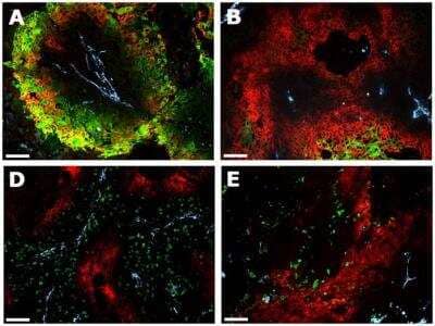
Immunocytochemistry/ Immunofluorescence: Carbonic Anhydrase IX/CA9 Antibody - BSA Free [NB100-417] - Immunofluorescent images showing CAIX expression, pimonidazole, apoptosis & proliferation in SCCNij202.CAIX is expressed in hypoxic regions as assessed by pimonidazole staining (A), but is also observed in non-pimonidazole areas (B). Yet, most of CAIX is expressed in pimonidazole positive regions (C). Proliferation (BrdUrd) & apoptosis (caspase-3) in relation to CAIX expression are shown in figure D & E respectively. Red, CAIX; Green, pimonidazole (A–B), BrdUrd (D) or caspase-3 (E); Yellow, overlap of CAIX (red) & pimonidazole (green); Light blue, vessels. Magnification 100×. Scale bars represent 100 µm. Closed circles represent CAIX expression in pimonidazole positive regions; open circles represent CAIX expression in pimonidazole negative regions. Abbrevations: 1× S4, 8 h, one i.p. injection S4, harvest after 8 hours; 1× S4, 24 h, one i.p. injection S4, harvest after 24 hours; 3× S4, one i.p. injection S4 a day for 3 days, harvest 8 hours after the last injection; 5× S4, one i.p. injection S4 a day for 5 days, harvest 8 hours after the last injection. Image collected & cropped by CiteAb from the following publication (https://pubmed.ncbi.nlm.nih.gov/25225880), licensed under a CC-BY license. Not internally tested by Novus Biologicals.
Immunohistochemistry: Carbonic Anhydrase IX/CA9 Antibody - BSA Free [NB100-417] -
Immunohistochemistry: Carbonic Anhydrase IX/CA9 Antibody - BSA Free [NB100-417] - Colorectal cancer xenograft growth was suppressed by CB-PIC(20 & 50 mg/kg body weight) in female athymic nude mice. Starting three days after SW620 cell inoculation, CB-PIC (20 & 50 mg/kg body weight) was injected in abdomen with 4% Tween 20 as vehicle once daily. (a) Body weights of mice. (b) Tumor growth in a time course. (c) Final tumor weight at termination of experiment. (d) Representative examples of immunohistochemical staining for Ki-67, CD34, TUNEL, CA IX & pAMPK alpha in tumor sections. Graphs show the Ki67 index (proliferation), CD34 index (angiogenesis), TUNEL index (apoptosis), CA IX (hypoxia region), & pAMPK alpha index in tumor sections. Values are means ± SD, n = 6. *P < 0.05 & ***P < 0.001 compared with control mice. Image collected & cropped by CiteAb from the following publication (https://pubmed.ncbi.nlm.nih.gov/23589723), licensed under a CC-BY license. Not internally tested by Novus Biologicals.
Western Blot: Carbonic Anhydrase IX/CA9 Antibody - BSA Free [NB100-417] -
Western Blot: Carbonic Anhydrase IX/CA9 Antibody - BSA Free [NB100-417] - Hypoxia induces GPRC5A directly via HIFsASILAC‐based proteomics data identify known (red) & novel (green) hypoxia‐induced proteins in SW620 cells. One‐sample t‐test was performed.BWestern blotting confirmed GPRC5A as a hypoxia‐induced protein in SILAC lysates.CValidation of GPRC5A Western blot data using siRNA. *Non‐specific band of ˜60 kDa not depleted by GPRC5A siRNA.DConfocal microscopy showing plasma membrane GPRC5A expression in hypoxic SW620 cells (scale bars: 75 μm).EWestern blotting showing GPRC5A upregulation by hypoxia in a panel of colorectal tumour cell lines.FBasal & hypoxia‐induced GPRC5A protein expression was decreased by HIF‐1/2 alpha depletion.GDepletion of HIF‐1 beta decreased GPRC5A protein upregulation in hypoxia.HHypoxia mimetic DMOG induced HIF‐1/2 alpha, CA9 & GPRC5A protein expression. Dual HIF‐1/2 alpha depletion reduced GPRC5A induction by DMOG.IqRT–PCR demonstrating that GPRC5A mRNA was upregulated by hypoxia (n = 3). GPRC5A was normalised to HPRT (error bars ± SD).JqRT–PCR demonstrating that HIF‐1/2 alpha depletion decreased GPRC5A induction during hypoxia (n = 3). GPRC5A was normalised to HPRT (error bars ± SD).KChIP‐PCR analyses identify HIF‐1 alpha binding to the GPRC5A promoter region containing a putative optimal HRE (error bars ± SD, n = 3).Data information: Asterisks (*) indicate non‐specific band. Level adjustments were made to images in Adobe Photoshop post‐acquisition for clarity (equal changes applied to the entire image). Representative examples of n = 3 independent experiments are shown.Source data are available online for this figure. Image collected & cropped by CiteAb from the following publication (https://pubmed.ncbi.nlm.nih.gov/30143543), licensed under a CC-BY license. Not internally tested by Novus Biologicals.
Immunocytochemistry/ Immunofluorescence: Carbonic Anhydrase IX/CA9 Antibody - BSA Free [NB100-417] -
Immunocytochemistry/ Immunofluorescence: Carbonic Anhydrase IX/CA9 Antibody - BSA Free [NB100-417] - Immunofluorescent images showing CAIX expression, pimonidazole, apoptosis & proliferation in SCCNij202.CAIX is expressed in hypoxic regions as assessed by pimonidazole staining (A), but is also observed in non-pimonidazole areas (B). Yet, most of CAIX is expressed in pimonidazole positive regions (C). Proliferation (BrdUrd) & apoptosis (caspase-3) in relation to CAIX expression are shown in figure D & E respectively. Red, CAIX; Green, pimonidazole (A–B), BrdUrd (D) or caspase-3 (E); Yellow, overlap of CAIX (red) & pimonidazole (green); Light blue, vessels. Magnification 100×. Scale bars represent 100 µm. Closed circles represent CAIX expression in pimonidazole positive regions; open circles represent CAIX expression in pimonidazole negative regions. Abbrevations: 1× S4, 8 h, one i.p. injection S4, harvest after 8 hours; 1× S4, 24 h, one i.p. injection S4, harvest after 24 hours; 3× S4, one i.p. injection S4 a day for 3 days, harvest 8 hours after the last injection; 5× S4, one i.p. injection S4 a day for 5 days, harvest 8 hours after the last injection. Image collected & cropped by CiteAb from the following publication (https://pubmed.ncbi.nlm.nih.gov/25225880), licensed under a CC-BY license. Not internally tested by Novus Biologicals.
Western Blot: Carbonic Anhydrase IX/CA9 Antibody - BSA Free [NB100-417] -
Western Blot: Carbonic Anhydrase IX/CA9 Antibody - BSA Free [NB100-417] - Establishment of a human protein replacement stable cell line using 47snoMEN.(a) Images of protein replacement stable cell lines (U2OSGFP–HIF-1 alpha-47PR). Expression of FP proteins was confirmed by fluorescence imaging with DFX treatment. Bar is 10 μm. (b) U2OS & U2OSGFP–HIF-1 alpha-47PR cells were exposed to 1% O2 for the indicated periods of time prior to lysis. Cell extracts were analysed by western blot using the indicated antibodies. (c) U2OS cells were transfected with 1 μg of control & HIF-1 alpha -47snoMEN vectors for 48 hours prior to lysis. DFX was added for the last 24 hours. Cell extracts were analysed by western blot using the indicated antibodies. (d) 3D culture analysis. HeLaGFP–HIF-1 alpha-47PR cells were cultured in lipidure coated plates to induce spheroid formation. HIF-1 alpha is visible in the core of the spheroid revealing hypoxia. Bar is 10 μm. Image collected & cropped by CiteAb from the following publication (https://dx.plos.org/10.1371/journal.pone.0154759), licensed under a CC-BY license. Not internally tested by Novus Biologicals.
Immunocytochemistry/ Immunofluorescence: Carbonic Anhydrase IX/CA9 Antibody - BSA Free [NB100-417] -
Immunocytochemistry/ Immunofluorescence: Carbonic Anhydrase IX/CA9 Antibody - BSA Free [NB100-417] - Immunofluorescent images showing CAIX expression, pimonidazole, apoptosis & proliferation in SCCNij202.CAIX is expressed in hypoxic regions as assessed by pimonidazole staining (A), but is also observed in non-pimonidazole areas (B). Yet, most of CAIX is expressed in pimonidazole positive regions (C). Proliferation (BrdUrd) & apoptosis (caspase-3) in relation to CAIX expression are shown in figure D & E respectively. Red, CAIX; Green, pimonidazole (A–B), BrdUrd (D) or caspase-3 (E); Yellow, overlap of CAIX (red) & pimonidazole (green); Light blue, vessels. Magnification 100×. Scale bars represent 100 µm. Closed circles represent CAIX expression in pimonidazole positive regions; open circles represent CAIX expression in pimonidazole negative regions. Abbrevations: 1× S4, 8 h, one i.p. injection S4, harvest after 8 hours; 1× S4, 24 h, one i.p. injection S4, harvest after 24 hours; 3× S4, one i.p. injection S4 a day for 3 days, harvest 8 hours after the last injection; 5× S4, one i.p. injection S4 a day for 5 days, harvest 8 hours after the last injection. Image collected & cropped by CiteAb from the following publication (https://pubmed.ncbi.nlm.nih.gov/25225880), licensed under a CC-BY license. Not internally tested by Novus Biologicals.



![Immunohistochemistry: Carbonic Anhydrase IX/CA9 Antibody - BSA Free [NB100-417] - Carbonic Anhydrase IX/CA9 Antibody - BSA Free](https://resources.bio-techne.com/images/products/nb100-417_rabbit-polyclonal-carbonic-anhydrase-ix-ca9-antibody-271220231253371.jpg)
![Immunohistochemistry: Carbonic Anhydrase IX/CA9 Antibody - BSA Free [NB100-417] - Carbonic Anhydrase IX/CA9 Antibody - BSA Free](https://resources.bio-techne.com/images/products/nb100-417_rabbit-polyclonal-carbonic-anhydrase-ix-ca9-antibody-271220231253316.jpg)
![Immunohistochemistry: Carbonic Anhydrase IX/CA9 Antibody - BSA Free [NB100-417] - Carbonic Anhydrase IX/CA9 Antibody - BSA Free](https://resources.bio-techne.com/images/products/nb100-417_rabbit-polyclonal-carbonic-anhydrase-ix-ca9-antibody-27122023128561.jpg)
![Western Blot: Carbonic Anhydrase IX/CA9 Antibody - BSA Free [NB100-417] - Carbonic Anhydrase IX/CA9 Antibody - BSA Free](https://resources.bio-techne.com/images/products/nb100-417_rabbit-polyclonal-carbonic-anhydrase-ix-ca9-antibody-310202415394513.jpg)
![Immunohistochemistry-Paraffin: Carbonic Anhydrase IX/CA9 Antibody - BSA Free [NB100-417] - Carbonic Anhydrase IX/CA9 Antibody - BSA Free](https://resources.bio-techne.com/images/products/nb100-417_rabbit-polyclonal-carbonic-anhydrase-ix-ca9-antibody-310202415382445.jpg)
![Immunohistochemistry: Carbonic Anhydrase IX/CA9 Antibody - BSA Free [NB100-417] - Carbonic Anhydrase IX/CA9 Antibody - BSA Free](https://resources.bio-techne.com/images/products/nb100-417_rabbit-polyclonal-carbonic-anhydrase-ix-ca9-antibody-310202415293313.jpg)
![Immunohistochemistry: Carbonic Anhydrase IX/CA9 Antibody - BSA Free [NB100-417] - Carbonic Anhydrase IX/CA9 Antibody - BSA Free](https://resources.bio-techne.com/images/products/nb100-417_rabbit-polyclonal-carbonic-anhydrase-ix-ca9-antibody-310202415171346.jpg)
![Immunohistochemistry: Carbonic Anhydrase IX/CA9 Antibody - BSA Free [NB100-417] - Carbonic Anhydrase IX/CA9 Antibody - BSA Free](https://resources.bio-techne.com/images/products/nb100-417_rabbit-polyclonal-carbonic-anhydrase-ix-ca9-antibody-310202415371932.jpg)
![Immunohistochemistry: Carbonic Anhydrase IX/CA9 Antibody - BSA Free [NB100-417] - Carbonic Anhydrase IX/CA9 Antibody - BSA Free](https://resources.bio-techne.com/images/products/nb100-417_rabbit-polyclonal-carbonic-anhydrase-ix-ca9-antibody-310202415304258.jpg)
![Western Blot: Carbonic Anhydrase IX/CA9 Antibody - BSA Free [NB100-417] - Carbonic Anhydrase IX/CA9 Antibody - BSA Free](https://resources.bio-techne.com/images/products/nb100-417_rabbit-polyclonal-carbonic-anhydrase-ix-ca9-antibody-31020241535610.jpg)
![Immunohistochemistry: Carbonic Anhydrase IX/CA9 Antibody - BSA Free [NB100-417] - Carbonic Anhydrase IX/CA9 Antibody - BSA Free](https://resources.bio-techne.com/images/products/nb100-417_rabbit-polyclonal-carbonic-anhydrase-ix-ca9-antibody-31020241529339.jpg)
![Western Blot: Carbonic Anhydrase IX/CA9 Antibody - BSA Free [NB100-417] - Carbonic Anhydrase IX/CA9 Antibody - BSA Free](https://resources.bio-techne.com/images/products/nb100-417_rabbit-polyclonal-carbonic-anhydrase-ix-ca9-antibody-31020241535621.jpg)
![Immunohistochemistry: Carbonic Anhydrase IX/CA9 Antibody - BSA Free [NB100-417] - Carbonic Anhydrase IX/CA9 Antibody - BSA Free](https://resources.bio-techne.com/images/products/nb100-417_rabbit-polyclonal-carbonic-anhydrase-ix-ca9-antibody-310202415392514.jpg)
![Immunohistochemistry: Carbonic Anhydrase IX/CA9 Antibody - BSA Free [NB100-417] - Carbonic Anhydrase IX/CA9 Antibody - BSA Free](https://resources.bio-techne.com/images/products/nb100-417_rabbit-polyclonal-carbonic-anhydrase-ix-ca9-antibody-310202415394539.jpg)
![Western Blot: Carbonic Anhydrase IX/CA9 Antibody - BSA Free [NB100-417] - Carbonic Anhydrase IX/CA9 Antibody - BSA Free](https://resources.bio-techne.com/images/products/nb100-417_rabbit-polyclonal-carbonic-anhydrase-ix-ca9-antibody-31020241539596.jpg)
![Immunocytochemistry/ Immunofluorescence: Carbonic Anhydrase IX/CA9 Antibody - BSA Free [NB100-417] - Carbonic Anhydrase IX/CA9 Antibody - BSA Free](https://resources.bio-techne.com/images/products/nb100-417_rabbit-polyclonal-carbonic-anhydrase-ix-ca9-antibody-310202415171339.jpg)
![Western Blot: Carbonic Anhydrase IX/CA9 Antibody - BSA Free [NB100-417] - Carbonic Anhydrase IX/CA9 Antibody - BSA Free](https://resources.bio-techne.com/images/products/nb100-417_rabbit-polyclonal-carbonic-anhydrase-ix-ca9-antibody-31020241612652.jpg)
![Immunohistochemistry: Carbonic Anhydrase IX/CA9 Antibody - BSA Free [NB100-417] - Carbonic Anhydrase IX/CA9 Antibody - BSA Free](https://resources.bio-techne.com/images/products/nb100-417_rabbit-polyclonal-carbonic-anhydrase-ix-ca9-antibody-310202415535129.jpg)
![Immunocytochemistry/ Immunofluorescence: Carbonic Anhydrase IX/CA9 Antibody - BSA Free [NB100-417] - Carbonic Anhydrase IX/CA9 Antibody - BSA Free](https://resources.bio-techne.com/images/products/nb100-417_rabbit-polyclonal-carbonic-anhydrase-ix-ca9-antibody-310202415525275.jpg)
![Immunocytochemistry/ Immunofluorescence: Carbonic Anhydrase IX/CA9 Antibody - BSA Free [NB100-417] - Carbonic Anhydrase IX/CA9 Antibody - BSA Free](https://resources.bio-techne.com/images/products/nb100-417_rabbit-polyclonal-carbonic-anhydrase-ix-ca9-antibody-31020241554518.jpg)
![Immunohistochemistry: Carbonic Anhydrase IX/CA9 Antibody - BSA Free [NB100-417] - Carbonic Anhydrase IX/CA9 Antibody - BSA Free](https://resources.bio-techne.com/images/products/nb100-417_rabbit-polyclonal-carbonic-anhydrase-ix-ca9-antibody-310202415525266.jpg)
![Western Blot: Carbonic Anhydrase IX/CA9 Antibody - BSA Free [NB100-417] - Carbonic Anhydrase IX/CA9 Antibody - BSA Free](https://resources.bio-techne.com/images/products/nb100-417_rabbit-polyclonal-carbonic-anhydrase-ix-ca9-antibody-310202415535151.jpg)
![Immunocytochemistry/ Immunofluorescence: Carbonic Anhydrase IX/CA9 Antibody - BSA Free [NB100-417] - Carbonic Anhydrase IX/CA9 Antibody - BSA Free](https://resources.bio-techne.com/images/products/nb100-417_rabbit-polyclonal-carbonic-anhydrase-ix-ca9-antibody-310202415525233.jpg)
![Immunocytochemistry/ Immunofluorescence: Carbonic Anhydrase IX/CA9 Antibody - BSA Free [NB100-417] - Carbonic Anhydrase IX/CA9 Antibody - BSA Free](https://resources.bio-techne.com/images/products/nb100-417_rabbit-polyclonal-carbonic-anhydrase-ix-ca9-antibody-31020241683941.jpg)
![Western Blot: Carbonic Anhydrase IX/CA9 Antibody - BSA Free [NB100-417] - Carbonic Anhydrase IX/CA9 Antibody - BSA Free](https://resources.bio-techne.com/images/products/nb100-417_rabbit-polyclonal-carbonic-anhydrase-ix-ca9-antibody-310202415541935.jpg)
![Immunocytochemistry/ Immunofluorescence: Carbonic Anhydrase IX/CA9 Antibody - BSA Free [NB100-417] - Carbonic Anhydrase IX/CA9 Antibody - BSA Free](https://resources.bio-techne.com/images/products/nb100-417_rabbit-polyclonal-carbonic-anhydrase-ix-ca9-antibody-31020241612665.jpg)