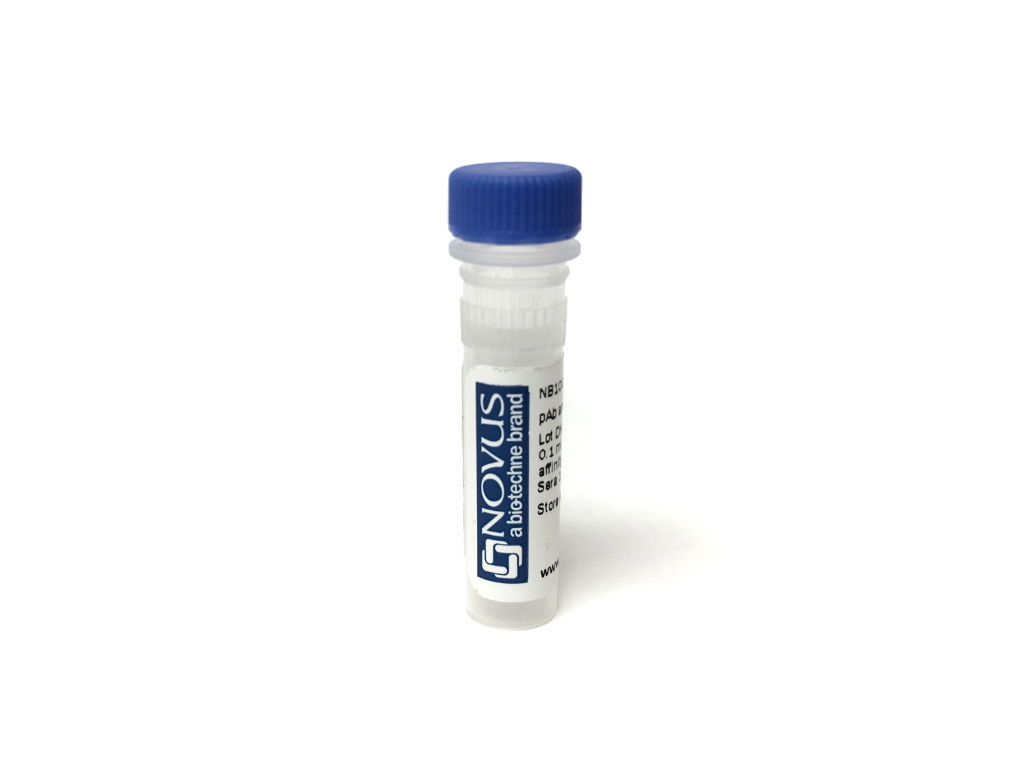
CD1.1 antigen Antibody (CB3) [CoraFluor™ 1]
Novus Biologicals, part of Bio-Techne | Catalog # NBP1-28362CL1


Conjugate
Catalog #
Forumulation
Catalog #
Key Product Details
Species Reactivity
Chicken
Applications
Flow Cytometry
Label
CoraFluor 1
Antibody Source
Monoclonal Mouse IgG1 kappa Clone # CB3
Concentration
Please see the vial label for concentration. If unlisted please contact technical services.
Product Specifications
Immunogen
Lymphocytes from the bursa of Fabricius of outbred chickens
Specificity
Chicken CD1.1
Clonality
Monoclonal
Host
Mouse
Isotype
IgG1 kappa
Description
CoraFluor(TM) 1 is a high performance terbium-based TR-FRET (Time-Resolved Fluorescence Resonance Energy Transfer) or TRF (Time-Resolved Fluorescence) donor for high throughput assay development. CoraFluor(IM) 1 absorbs UV light at approximately 340 nm, and emits at approximately 490 nm, 545 nm, 585 nm and 620 nm. It is compatible with common acceptor dyes that absorb at the emission wavelengths of CoraFluor(TM) 1. CoraFluor(TM) 1 can be used for the development of robust and scalable TR-FRET binding assays such as target engagement, ternary complex, protein-protein interaction and protein quantification assays.
Applications for CD1.1 antigen Antibody (CB3) [CoraFluor™ 1]
Application
Recommended Usage
Flow Cytometry
Optimal dilutions of this antibody should be experimentally determined.
Application Notes
Optimal dilution of this antibody should be experimentally determined.
Formulation, Preparation, and Storage
Purification
Protein A or G purified
Formulation
PBS
Preservative
No Preservative
Concentration
Please see the vial label for concentration. If unlisted please contact technical services.
Shipping
The product is shipped with polar packs. Upon receipt, store it immediately at the temperature recommended below.
Stability & Storage
Store at 4C in the dark. Do not freeze.
Background: CD1.1 antigen
Alternate Names
CD1, CD1a antigen, CD1A antigen, a polypeptide, CD1a molecule, cluster of differentiation 1 A, cortical thymocyte antigen CD1A, differentiation antigen CD1-alpha-3, epidermal dendritic cell marker CD1a, FCB6, HTA1, hTa1 thymocyte antigen, R4, T6, T-cell surface antigen T6/Leu-6, T-cell surface glycoprotein CD1a
Gene Symbol
CD1A
Additional CD1.1 antigen Products
Product Documents for CD1.1 antigen Antibody (CB3) [CoraFluor™ 1]
Product Specific Notices for CD1.1 antigen Antibody (CB3) [CoraFluor™ 1]
CoraFluor (TM) is a trademark of Bio-Techne Corp. Sold for research purposes only under agreement from Massachusetts General Hospital. US patent 2022/0025254
This product is for research use only and is not approved for use in humans or in clinical diagnosis. Primary Antibodies are guaranteed for 1 year from date of receipt.
Loading...
Loading...
Loading...
Loading...
Loading...
Loading...