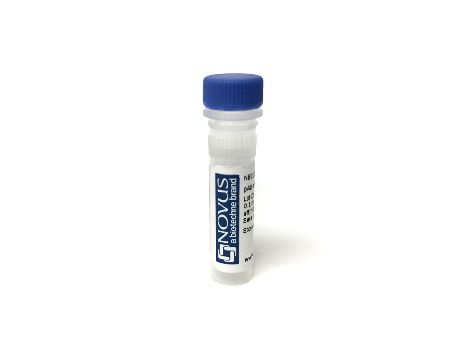
CD27/TNFRSF7 Antibody (57703) [PE/Atto594]
Novus Biologicals, part of Bio-Techne | Catalog # FAB382PEATT594


Conjugate
Catalog #
Key Product Details
Species Reactivity
Human
Applications
Flow Cytometry
Label
PE/Atto594 (Excitation = 488 nm, Emission = 627 nm)
Antibody Source
Monoclonal Mouse IgG1 Clone # 57703
Concentration
Please see the vial label for concentration. If unlisted please contact technical services.
Product Specifications
Immunogen
S. frugiperda insect ovarian cell line Sf 21-derived recombinant human CD27
Thr21-Ile192
Accession # P26842
Thr21-Ile192
Accession # P26842
Specificity
Detects human CD27 in direct ELISAs and Western blots. In direct ELISAs and Western blots, no cross‑reactivity with recombinant human (rh) 4‑1BB, rhBAFF R, recombinant mouse (rm) CD27, rhCD30, rhCD40, rhDR3, rhDR6, rhEDAR, rhFas, rhGITR, rhHVEM, rhLTR beta, rhNGF R, rhOPG, rmOX40, rhRANK, rhTAJ, or rhTNF RI is observed.
Clonality
Monoclonal
Host
Mouse
Isotype
IgG1
Applications for CD27/TNFRSF7 Antibody (57703) [PE/Atto594]
Application
Recommended Usage
Flow Cytometry
Optimal dilutions of this antibody should be experimentally determined.
Application Notes
Optimal dilution of this antibody should be experimentally determined. For optimal results using our Tandem dyes, please avoid prolonged exposure to light or extreme temperature fluctuations. These can lead to irreversible degradation or decoupling. When staining intracellular targets, specific attention to the fixation and permeabilization steps in your flow protocol may be required. Please contact our technical support team at technical@novusbio.com if you have any questions.
Formulation, Preparation, and Storage
Purification
Protein A or G purified from hybridoma culture supernatant
Formulation
PBS
Preservative
0.05% Sodium Azide
Concentration
Please see the vial label for concentration. If unlisted please contact technical services.
Shipping
The product is shipped with polar packs. Upon receipt, store it immediately at the temperature recommended below.
Stability & Storage
Store at 4C in the dark. Do not freeze.
Background: CD27/TNFRSF7
Membrane-bound CD27 is expressed as a disulfide-linked homodimer (3). CD27 binds to the ligand CD70, a transmembrane glycoprotein that is transiently expressed on activated immune cells such as antigen presenting cells (APCs), dendritic cells (DCs), NK cells, B cells, and T cells (1,2,6,7). The receptor-ligand binding interaction leads to NFkappaB and c-Jun pathway activation which promotes immune stimulation and activation and survival of CD4+ T cells, CD8+ T cells, memory T cells, and NK cells (2,6,7). Both CD27 and CD70 are often abnormally expressed or dysregulated on malignant and cancer cells leading to immune evasion and tumor progression (7). CD27 has become a target of interest of immunotherapies for viral infections, autoimmune disease, and cancer (2). Varlilumab, an agonistic CD27 monoclonal antibody (mAB), has entered clinical trials for the treatment of hematological and solid tumor cancers (1,6). Additional clinical trials are in process that combine varlilumab with other immune checkpoint inhibitors like the programmed cell death protein-1 (PD-1) blocking mAb nivolumab (1,2). Initial results are promising, suggesting that targeting CD27, especially in combination with other therapeutics, may be a promising and effective immunotherapy for a variety of pathologies (1,2,6).
References
1. Starzer AM, Berghoff AS. New emerging targets in cancer immunotherapy: CD27 (TNFRSF7). ESMO Open. 2020;4(Suppl 3):e000629. https://doi.org/10.1136/esmoopen-2019-000629
2. Grant EJ, Nussing S, Sant S, Clemens EB, Kedzierska K. The role of CD27 in anti-viral T-cell immunity. Curr Opin Virol. 2017;22:77-88. https://doi.org/10.1016/j.coviro.2016.12.001
3. Buchan SL, Rogel A, Al-Shamkhani A. The immunobiology of CD27 and OX40 and their potential as targets for cancer immunotherapy. Blood. 2018;131(1):39-48. https://10.1182/blood-2017-07-741025
4. Uniprot (P26842)
5. Uniprot (P41272)
6. van de Ven K, Borst J. Targeting the T-cell co-stimulatory CD27/CD70 pathway in cancer immunotherapy: rationale and potential. Immunotherapy. 2015;7(6):655-667. https://doi.org/10.2217/imt.15.32
7. Flieswasser T, Van den Eynde A, Van Audenaerde J, et al. The CD70-CD27 axis in oncology: the new kids on the block. J Exp Clin Cancer Res. 2022;41(1):12. https://doi.org/10.1186/s13046-021-02215-y
Alternate Names
CD27, TNFRSF7
Gene Symbol
CD27
Additional CD27/TNFRSF7 Products
Product Documents for CD27/TNFRSF7 Antibody (57703) [PE/Atto594]
Product Specific Notices for CD27/TNFRSF7 Antibody (57703) [PE/Atto594]
This product is for research use only and is not approved for use in humans or in clinical diagnosis. Primary Antibodies are guaranteed for 1 year from date of receipt.
Loading...
Loading...
Loading...
Loading...
Loading...
Loading...