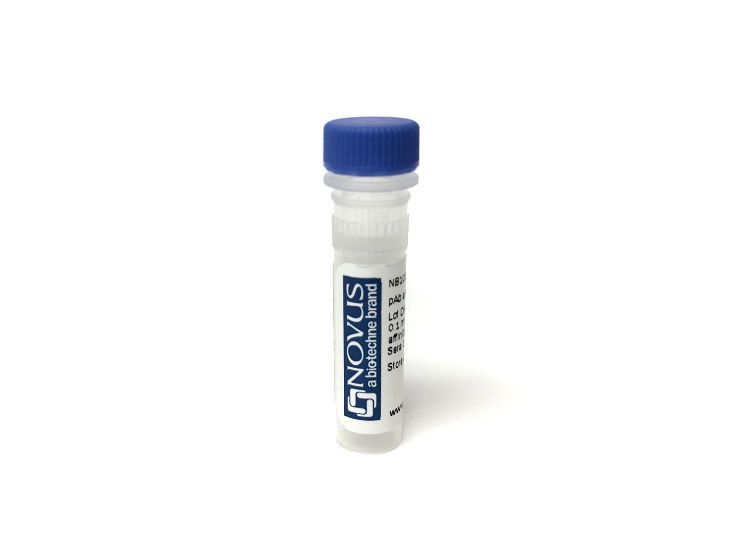
CD3 Antibody (17A2) [mFluor Violet 450 SE]
Novus Biologicals, part of Bio-Techne | Catalog # FAB4841MFV450


Conjugate
Catalog #
Key Product Details
Species Reactivity
Mouse
Applications
Complement-dependent Cytotoxicity, CyTOF-ready, Flow Cytometry, Immunocytochemistry, Immunohistochemistry, Immunoprecipitation, T Cell Stimulation
Label
mFluor Violet 450 SE (Excitation = 406 nm, Emission = 445 nm)
Antibody Source
Monoclonal Rat IgG2B Clone # 17A2
Concentration
Please see the vial label for concentration. If unlisted please contact technical services.
Product Specifications
Immunogen
T cell hybridoma D1
Specificity
Reacts with mouse TCR-associated CD3 complex that occurs on thymocytes and mature T cells.
Clonality
Monoclonal
Host
Rat
Isotype
IgG2B
Applications for CD3 Antibody (17A2) [mFluor Violet 450 SE]
Application
Recommended Usage
Complement-dependent Cytotoxicity
Optimal dilutions of this antibody should be experimentally determined.
CyTOF-ready
Optimal dilutions of this antibody should be experimentally determined.
Flow Cytometry
Optimal dilutions of this antibody should be experimentally determined.
Immunocytochemistry
Optimal dilutions of this antibody should be experimentally determined.
Immunohistochemistry
Optimal dilutions of this antibody should be experimentally determined.
Immunoprecipitation
Optimal dilutions of this antibody should be experimentally determined.
T Cell Stimulation
Optimal dilutions of this antibody should be experimentally determined.
Application Notes
Optimal dilution of this antibody should be experimentally determined.
Formulation, Preparation, and Storage
Purification
0
Formulation
50mM Sodium Borate
Preservative
0.05% Sodium Azide
Concentration
Please see the vial label for concentration. If unlisted please contact technical services.
Shipping
The product is shipped with polar packs. Upon receipt, store it immediately at the temperature recommended below.
Stability & Storage
Store at 4C in the dark.
Background: CD3
CD3 proteins are expressed on the surface of thymocytes during thymocyte development, proliferation, and maturation to T-cells (4, 6, 7). During T-cell development CD4-CD8- double negative (DN) cells differentiate to CD4+CD8+ double positive (DP) cells before progressing to single positive (SP) CD4+ helper T-cells or CD8+ cytotoxic T-cells (4, 6, 7). As CD3 plays an important role in thymocyte development, it is understandable that CD3 defects and mutations in CD3 protein chains cause severe combined immunodeficiencies (SCIDs) (8). Additionally, a subset of CD3+ T-cells that co-express CD20 are described in a variety of diseases including rheumatoid arthritis, multiple sclerosis, CD20+ T-cell leukemia/lymphoma, and HIV (9). Clinical trials and animal models have shown that anti-CD3 monoclonal antibodies are a promising treatment modality for inflammatory disorders and autoimmune diseases, such as type I diabetes (10).
References
1. Chetty, R., & Gatter, K. (1994). CD3: structure, function, and role of immunostaining in clinical practice. The Journal of pathology. https://doi.org/10.1002/path.1711730404
2. Mariuzza, R. A., Agnihotri, P., & Orban, J. (2020). The structural basis of T-cell receptor (TCR) activation: An enduring enigma. The Journal of biological chemistry. https://doi.org/10.1074/jbc.REV119.009411
3. Kuhns, M. S., Davis, M. M., & Garcia, K. C. (2006). Deconstructing the form and function of the TCR/CD3 complex. Immunity. https://doi.org/10.1016/j.immuni.2006.01.006
4. Clevers, H., Alarcon, B., Wileman, T., & Terhorst, C. (1988). The T cell receptor/CD3 complex: a dynamic protein ensemble. Annual review of immunology. https://doi.org/10.1146/annurev.iy.06.040188.003213
5. Uniprot: CD3-delta (P04234), CD3-epsilon (P07766), CD3-gamma (P09693), CD3-zeta (P20963)
6. D'Acquisto, F., & Crompton, T. (2011). CD3+CD4-CD8- (double negative) T cells: saviours or villains of the immune response?. Biochemical pharmacology. https://doi.org/10.1016/j.bcp.2011.05.019
7. Dave V. P. (2009). Hierarchical role of CD3 chains in thymocyte development. Immunological reviews. https://doi.org/10.1111/j.1600-065X.2009.00835.x
8. Fischer, A., de Saint Basile, G., & Le Deist, F. (2005). CD3 deficiencies. Current opinion in allergy and clinical immunology. https://doi.org/10.1097/01.all.0000191886.12645.79
9. Chen, Q., Yuan, S., Sun, H., & Peng, L. (2019). CD3+CD20+ T cells and their roles in human diseases. Human immunology. https://doi.org/10.1016/j.humimm.2019.01.001
10. Kuhn, C., & Weiner, H. L. (2016). Therapeutic anti-CD3 monoclonal antibodies: from bench to bedside. Immunotherapy. https://doi.org/10.2217/imt-2016-0049
Alternate Names
CD_antigen: CD3e, CD3 antigen, delta subunit, CD3d antigen, CD3d antigen, delta polypeptide (TiT3 complex), CD3d molecule, delta (CD3-TCR complex), CD3-DELTA, CD3e, CD3e antigen, CD3e antigen, epsilon polypeptide (TiT3 complex), CD3e molecule, epsilon (CD3-TCR complex), CD3-epsilon, CD3G, CD3g antigen, CD3g antigen, gamma polypeptide (TiT3 complex), CD3g molecule, epsilon (CD3-TCR complex), CD3g molecule, gamma (CD3-TCR complex), CD3-GAMMA, FLJ17620, FLJ17664, FLJ18683, FLJ79544, FLJ94613, IMD18, MGC138597, T3DOKT3, delta chain, T3E, T-cell antigen receptor complex, epsilon subunit of T3, T-cell receptor T3 delta chain, T-cell surface antigen T3/Leu-4 epsilon chain, T-cell surface glycoprotein CD3 delta chain, T-cell surface glycoprotein CD3 epsilon chain, TCRE
Gene Symbol
CD3E
Additional CD3 Products
Product Documents for CD3 Antibody (17A2) [mFluor Violet 450 SE]
Product Specific Notices for CD3 Antibody (17A2) [mFluor Violet 450 SE]
mFluor(TM) is a trademark of AAT Bioquest, Inc. This conjugate is made on demand. Actual recovery may vary from the stated volume of this product. The volume will be greater than or equal to the unit size stated on the datasheet.
This product is for research use only and is not approved for use in humans or in clinical diagnosis. Primary Antibodies are guaranteed for 1 year from date of receipt.
Loading...
Loading...
Loading...
Loading...
Loading...
Loading...