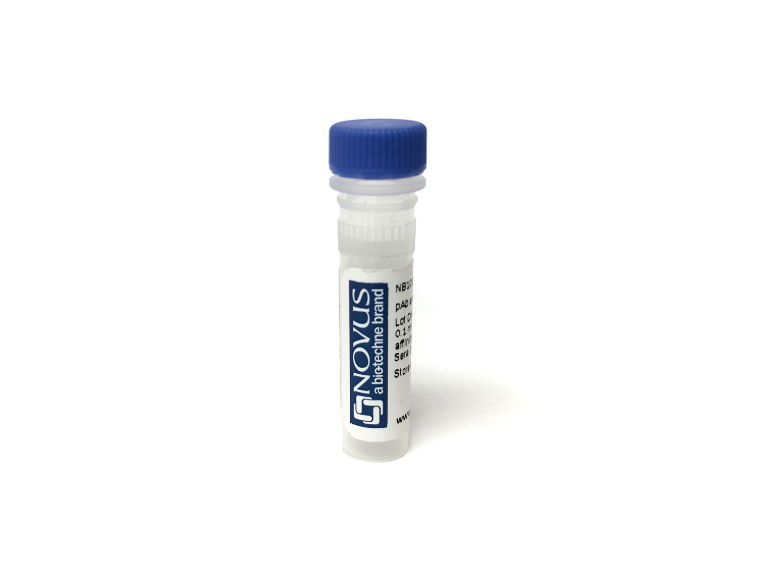
CD3 epsilon Antibody (PC3/188A) [Allophycocyanin/Cy7]
Novus Biologicals, part of Bio-Techne | Catalog # NBP2-54405APCCY7


Conjugate
Catalog #
Forumulation
Catalog #
Key Product Details
Species Reactivity
Human, Chimpanzee
Applications
CyTOF-ready, Flow Cytometry, Immunocytochemistry/ Immunofluorescence, Immunohistochemistry (Negative), Western Blot
Label
Allophycocyanin/Cy7 (Excitation = 650;755 nm, Emission = 767 nm)
Antibody Source
Monoclonal Mouse IgG1 Clone # PC3/188A
Concentration
Please see the vial label for concentration. If unlisted please contact technical services.
Product Specifications
Immunogen
A synthetic peptide corresponding to amino acids 156-168 of the cytoplasmic domain of human CD3- chain (Uniprot: P07766 )
Reactivity Notes
Others not known.
Localization
Cell surface and Cytoplasmic
Specificity
Recognizes the epsilon-chain of CD3, which consists of five different polypeptide chains (designated as gamma, delta, epsilon, zeta, and eta) with MW ranging from 16-28kDa. The CD3 complex is closely associated at the lymphocyte cell surface with the T cell antigen receptor (TCR). Reportedly, CD3 complex is involved in signal transduction to the T cell interior following antigen recognition. The CD3 antigen is first detectable in early thymocytes and probably represents one of the earliest signs of commitment to the T cell lineage. In cortical thymocytes, CD3 is predominantly intra-cytoplasmic. However, in medullary thymocytes, it appears on the T cell surface. CD3 antigen is a highly specific marker for T cells, and is present in majority of T cell neoplasms.
Marker
T-Cell Marker
Clonality
Monoclonal
Host
Mouse
Isotype
IgG1
Applications for CD3 epsilon Antibody (PC3/188A) [Allophycocyanin/Cy7]
Application
Recommended Usage
CyTOF-ready
Optimal dilutions of this antibody should be experimentally determined.
Flow Cytometry
Optimal dilutions of this antibody should be experimentally determined.
Immunocytochemistry/ Immunofluorescence
Optimal dilutions of this antibody should be experimentally determined.
Immunohistochemistry (Negative)
Optimal dilutions of this antibody should be experimentally determined.
Western Blot
Optimal dilutions of this antibody should be experimentally determined.
Application Notes
Optimal dilution of this antibody should be experimentally determined. For optimal results using our Tandem dyes, please avoid prolonged exposure to light or extreme temperature fluctuations. These can lead to irreversible degradation or decoupling. When staining intracellular targets, specific attention to the fixation and permeabilization steps in your flow protocol may be required. Please contact our technical support team at technical@novusbio.com if you have any questions.
Formulation, Preparation, and Storage
Purification
Protein A or G purified
Formulation
PBS
Preservative
0.05% Sodium Azide
Concentration
Please see the vial label for concentration. If unlisted please contact technical services.
Shipping
The product is shipped with polar packs. Upon receipt, store it immediately at the temperature recommended below.
Stability & Storage
Store at 4C in the dark. Do not freeze.
Background: CD3 epsilon
Alternate Names
CD3e
Gene Symbol
CD3E
Additional CD3 epsilon Products
Product Documents for CD3 epsilon Antibody (PC3/188A) [Allophycocyanin/Cy7]
Product Specific Notices for CD3 epsilon Antibody (PC3/188A) [Allophycocyanin/Cy7]
This product is for research use only and is not approved for use in humans or in clinical diagnosis. Primary Antibodies are guaranteed for 1 year from date of receipt.
Loading...
Loading...
Loading...
Loading...
Loading...
Loading...