CD31/PECAM-1 Antibody (JC/70A) - BSA Free
Novus Biologicals, part of Bio-Techne | Catalog # NB600-562
Clone JC/70A was used by HLDA to establish CD designation.

Conjugate
Catalog #
Forumulation
Catalog #
Key Product Details
Validated by
Knockout/Knockdown, Orthogonal Validation
Species Reactivity
Validated:
Human, Mouse, Feline, Rabbit
Cited:
Human, Mouse, Rat, Rabbit
Applications
Validated:
Dual RNAscope ISH-IHC, ELISA, Flow (Cell Surface), Flow Cytometry, Immunocytochemistry/ Immunofluorescence, Immunohistochemistry, Immunohistochemistry-Frozen, Immunohistochemistry-Paraffin, Knockout Validated, Western Blot
Cited:
ELISA, Flow Cytometry, IF/IHC, Immunocytochemistry/ Immunofluorescence, Immunohistochemistry, Immunohistochemistry Whole-Mount, Immunohistochemistry-Frozen, Immunohistochemistry-Paraffin, Western Blot
Label
Unconjugated
Antibody Source
Monoclonal Mouse IgG1 kappa Clone # JC/70A
Format
BSA Free
Concentration
1.0 mg/ml
Product Specifications
Immunogen
This CD31/PECAM-1 Antibody (JC/70A) was developed against a membrane preparation of a spleen from a patient with hairy cell leukemia.
Reactivity Notes
Rabbit (PMID: 21533193) and Mouse (PMID: 29700126) reactivity reported in scientific literature.
Localization
Cell membrane.
Clonality
Monoclonal
Host
Mouse
Isotype
IgG1 kappa
Theoretical MW
82.5 kDa.
Disclaimer note: The observed molecular weight of the protein may vary from the listed predicted molecular weight due to post translational modifications, post translation cleavages, relative charges, and other experimental factors.
Disclaimer note: The observed molecular weight of the protein may vary from the listed predicted molecular weight due to post translational modifications, post translation cleavages, relative charges, and other experimental factors.
Scientific Data Images for CD31/PECAM-1 Antibody (JC/70A) - BSA Free
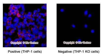
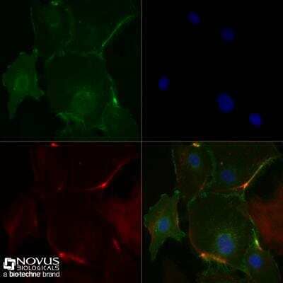
Knockout Validation of CD31/PECAM-1 Antibody in THP-1 Cells by Immunocytochemistry/Immunofluorescence
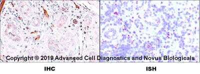
CD31/PECAM-1 was detected in immersion fixed THP-1 (red, positive) but not THP-1 KO (negative) cells using 8 ug/mL Mouse Anti-Human CD31/PECAM-1 (JC/70A) Monoclonal Antibody (Catalog # NB600-562) for 3 hours at room temperature. Cells were stained with the NorthernLights(TM) 557-conjugated Donkey anti-Mouse IgG Secondary Antibody (red, Catalog # NL007)) and and counterstained with DAPI (blue).Dual RNAscope ISH-IHC Analysis of CD31/PECAM-1 in Human Tonsil
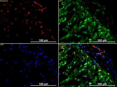
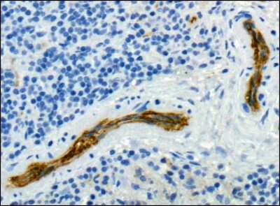
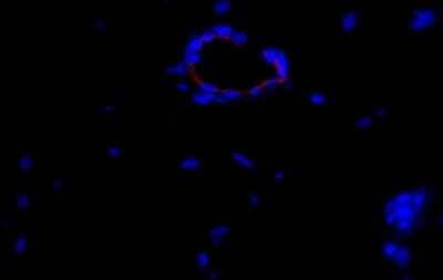
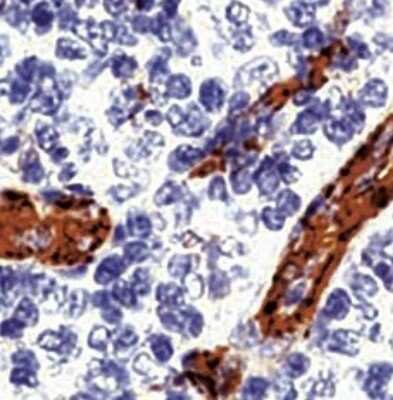
Formalin-fixed paraffin-embedded tissue sections of human metastatic tonsil were probed for CD31 mRNA (ACD RNAScope probe, catalog # 487381; Fast Red chromogen, ACD catalog # 322500). Adjacent tissue section was processes for immunohistochemistry using mouse monoclonal (NB600-562) at 1:25 dilution for 1 hour at room temperature followed by incubation with the anti-mouse IgG VisUCyte HRP Polymer Antibody (Catalog # VC001) and DAB chromogen (yellow-brown). Tissue was counterstained with hematoxylin (blue).Immunohistological Staining of CD31/PECAM-1 in Frozen Rabbit Heart
Rabbit Heart, CD31 Stained Red. Image from verified customer review.Applications for CD31/PECAM-1 Antibody (JC/70A) - BSA Free
Application
Recommended Usage
Flow (Cell Surface)
1:100-1:250
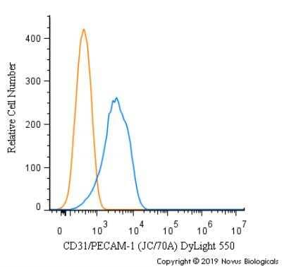
Flow Cytometry
1:100-1:250
Immunohistochemistry
1:10-1:25
Immunohistochemistry-Paraffin
1:10-1:25
Western Blot
1:100-1:500
Application Notes
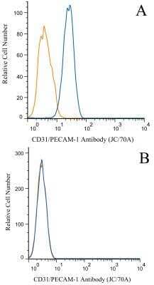
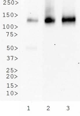
This CD31/PECAM1 Antibody (JC/70A) is useful for IHC on paraffin-embedded sections, Flow cytometry and Western blot. It reacts with an ~100 kDa glycoprotein expressed by endothelial cells and ~130 kDa glycoprotein present in platelets. It stains endothelial cells in normal as well as malignant tissues. Prior to immunostaining paraffin tissues, antigen retrieval with sodium citrate buffer (pH 6.0) is recommended. Use in ELISA reported in scientific literature (PMID: 8307606).
Reviewed Applications
Read 4 reviews rated 4.3 using NB600-562 in the following applications:
Formulation, Preparation, and Storage
Purification
Protein G purified
Formulation
PBS
Format
BSA Free
Preservative
0.02% Sodium Azide
Concentration
1.0 mg/ml
Shipping
The product is shipped with polar packs. Upon receipt, store it immediately at the temperature recommended below.
Stability & Storage
Store at 4C short term. Aliquot and store at -20C long term. Avoid freeze-thaw cycles. After opening store under sterile conditions.
Background: CD31/PECAM-1
PECAM's intracellular cytoplasmic domain consists of a sequence of 118 amino acids and contains serine and tyrosine (also referred to as immunoreceptor tyrosine-based inhibitory motifs-ITIMs) residues, which may be phosphorylated upon cellular stimulation (3). ITIMs are phosphorylated by Src-family kinases and non-Src family kinases (e.g., Csk), leading to a conformational change which supports interactions with Src homology 2 (SH2) domain containing proteins such as protein-tyrosine phosphatase, SHP-2 (1,2). Formation of SHP-2/PECAM-1 complexes induces endothelial cell migration through the dephosphorylation of focal adhesion kinase and regulation of RhoA activity (1). Signaling downstream of ITIM tyrosine phosphorylations also plays a role in PECAM's anti-apoptotic activity, a function which is independent of its interaction with SHP-2. In platelets and leukocytes, phosphorylation of PECAM's cytosolic domain is inhibitory, preventing their activation.
References
1. Lertkiatmongkol, P., Liao, D., Mei, H., Hu, Y., & Newman, P. J. (2016). Endothelial functions of PECAM-1 (CD31). Current Opinion in Hematology. https://doi.org/10.1097/MOH.0000000000000239.Endothelial
2. Privratsky, J. R., & Newman, P. J. (2014). PECAM-1: Regulator of endothelial junctional integrity. Cell and Tissue Research. https://doi.org/10.1007/s00441-013-1779-3
3. Newman, P. J., & Newman, D. K. (2003). Signal transduction pathways mediated by PECAM-1: New roles for an old molecule in platelet and vascular cell biology. Arteriosclerosis, Thrombosis, and Vascular Biology. https://doi.org/10.1161/01.ATV.0000071347.69358.D9
Long Name
Platelet Endothelial Cell Adhesion Molecule 1
Alternate Names
CD31, EndoCAM, PECA1, PECAM-1, PECAM1, JC70A CD31, JC70A Clone
Entrez Gene IDs
5175 (Human)
Gene Symbol
PECAM1
UniProt
Additional CD31/PECAM-1 Products
Product Documents for CD31/PECAM-1 Antibody (JC/70A) - BSA Free
Product Specific Notices for CD31/PECAM-1 Antibody (JC/70A) - BSA Free
This product is for research use only and is not approved for use in humans or in clinical diagnosis. Primary Antibodies are guaranteed for 1 year from date of receipt.
Loading...
Loading...
Loading...
Loading...
Loading...
Loading...

![Immunocytochemistry/ Immunofluorescence: CD31/PECAM-1 Antibody (JC/70A) - BSA Free [NB600-562] - CD31/PECAM-1 Antibody (JC/70A) - BSA Free](https://resources.bio-techne.com/images/products/nb600-562_mouse-monoclonal-cd31-pecam-1-antibody-jc-70a-310202416171426.jpg)
![Immunocytochemistry/ Immunofluorescence: CD31/PECAM-1 Antibody (JC/70A) - BSA Free [NB600-562] - CD31/PECAM-1 Antibody (JC/70A) - BSA Free](https://resources.bio-techne.com/images/products/nb600-562_mouse-monoclonal-cd31-pecam-1-antibody-jc-70a-31020241618941.jpg)
![Immunocytochemistry/ Immunofluorescence: CD31/PECAM-1 Antibody (JC/70A) - BSA Free [NB600-562] - CD31/PECAM-1 Antibody (JC/70A) - BSA Free](https://resources.bio-techne.com/images/products/nb600-562_mouse-monoclonal-cd31-pecam-1-antibody-jc-70a-310202416173525.jpg)
![Immunocytochemistry/ Immunofluorescence: CD31/PECAM-1 Antibody (JC/70A) - BSA Free [NB600-562] - CD31/PECAM-1 Antibody (JC/70A) - BSA Free](https://resources.bio-techne.com/images/products/nb600-562_mouse-monoclonal-cd31-pecam-1-antibody-jc-70a-31020241616378.jpg)
![Immunocytochemistry/ Immunofluorescence: CD31/PECAM-1 Antibody (JC/70A) - BSA Free [NB600-562] - CD31/PECAM-1 Antibody (JC/70A) - BSA Free](https://resources.bio-techne.com/images/products/nb600-562_mouse-monoclonal-cd31-pecam-1-antibody-jc-70a-3102024161609.jpg)
![Immunocytochemistry/ Immunofluorescence: CD31/PECAM-1 Antibody (JC/70A) - BSA Free [NB600-562] - CD31/PECAM-1 Antibody (JC/70A) - BSA Free](https://resources.bio-techne.com/images/products/nb600-562_mouse-monoclonal-cd31-pecam-1-antibody-jc-70a-310202416163753.jpg)
![Immunocytochemistry/ Immunofluorescence: CD31/PECAM-1 Antibody (JC/70A) - BSA Free [NB600-562] - CD31/PECAM-1 Antibody (JC/70A) - BSA Free](https://resources.bio-techne.com/images/products/nb600-562_mouse-monoclonal-cd31-pecam-1-antibody-jc-70a-310202416171483.jpg)