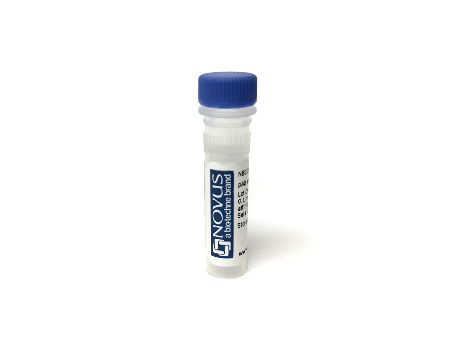
CD4 Antibody (CD4/8203R) [DyLight 650]
Novus Biologicals, part of Bio-Techne | Catalog # NBP3-20762C
Recombinant Monoclonal Antibody


Conjugate
Catalog #
Forumulation
Catalog #
Key Product Details
Species Reactivity
Human
Applications
Immunohistochemistry-Paraffin
Label
DyLight 650 (Excitation = 652 nm, Emission = 672 nm)
Antibody Source
Recombinant Monoclonal Rabbit IgG Kappa Clone # CD4/8203R
Concentration
Please see the vial label for concentration. If unlisted please contact technical services.
Product Specifications
Immunogen
Recombinant fragment (around aa 200-400) of human CD4 protein (exact sequence is proprietary)
Localization
Cell surface.
Specificity
Anti-CD4 is used in the immunohistochemical staining of lymphoproliferative disorders to evaluate tumors with CD4 aberrant expression
Clonality
Monoclonal
Host
Rabbit
Isotype
IgG Kappa
Applications for CD4 Antibody (CD4/8203R) [DyLight 650]
Application
Recommended Usage
Immunohistochemistry-Paraffin
Optimal dilutions of this antibody should be experimentally determined.
Application Notes
Optimal dilution of this antibody should be experimentally determined.
Formulation, Preparation, and Storage
Purification
Protein A or G purified
Formulation
50mM Sodium Borate
Preservative
0.05% Sodium Azide
Concentration
Please see the vial label for concentration. If unlisted please contact technical services.
Shipping
The product is shipped with polar packs. Upon receipt, store it immediately at the temperature recommended below.
Stability & Storage
Store at 4C in the dark.
Background: CD4
Given its critical role in T cell development, CD4 also has diverse immunology-related functions. CD4 acts as a coreceptor with the T-cell receptor (TCR) during T cell activation and thymic differentiation by binding directly to major histocompatibility complex (MHC) class II antigens and associating with the protein tyrosine kinase, Lck (4). This interaction contributes to the formation of the immunological synapse (5). Defects in antigen presentation cause dysfunction of CD4+ T cells and the almost complete loss of MHC II expression on B cells in peripheral blood, as observed in severe combined immunodeficiency (SCID) (6). CD4 also functions as a receptor for the human immunodeficiency virus (HIV) by binding to gp120, the envelope glycoprotein of HIV-1. It has been shown that the V-like domains are critical for binding to gp120 (7). In immune mediated and infectious diseases of the central nervous system, CD4 functions as an indirect mediator of neuronal damage (8).
References
1. Omri, B., Crisanti, P., Alliot, F., Marty, M., Rutin, J., Levallois, C., . . . Pessac, B. (1994). CD4 expression in neurons of the central nervous system. International Immunology, 6(3), 377-385. doi:10.1093/intimm/6.3.377
2. Wan, Y. Y., & Flavell, R. A. (2009). How diverse-CD4 effector T cells and their functions. Journal of Molecular Cell Biology, 1(1), 20-36. doi:10.1093/jmcb/mjp001
3. Wu, H., Myszka, D. G., Tendian, S. W., Brouillette, C. G., Sweet, R. W., Chaiken, I. M., & Hendrickson, W. A. (1996). Kinetic and structural analysis of mutant CD4 receptors that are defective in HIV gp120 binding. Proceedings of the National Academy of Sciences, 93(26), 15030-15035. doi:10.1073/pnas.93.26.15030
4. Doyle, C., & Strominger, J. L. (1987). Interaction between CD4 and class II MHC molecules mediates cell adhesion. Nature, 330, 256-259. doi:10.1038/330256a0
5. Vignali, D. A. (2010). CD4 on the road to coreceptor status. The Journal of Immunology, 184(11), 5933-5934. doi:10.4049/jimmunol.1090037
6. Tasher, D., & Dalal, I. (2012). The genetic basis of severe combined immunodeficiency and its variants. The Application of Clinical Genetics, 5, 67-80. doi:10.2147/tacg.s18693
7. Arthos, J., Deen, K. C., Chaikin, M. A., Fornwald, J. A., Sathe, G., Sattentau, Q. J., . . . Sweet, R. W. (1989). Identification of the residues in human CD4 critical for the binding of HIV. Cell, 57(3), 469-481. doi:10.1016/0092-8674(89)90922-7
8. Buttini, M., Westland, C. E., Masliah, E., Yafeh, A. M., Wyss-Coray, T., Mucke, L. (1998). Novel role of human cd4 molecule identified in neurodegeneration. Nature Medicine, 4(4), 441-446. doi:10.1038/nm0498-441
Alternate Names
CD4
Gene Symbol
CD4
Additional CD4 Products
Product Documents for CD4 Antibody (CD4/8203R) [DyLight 650]
Product Specific Notices for CD4 Antibody (CD4/8203R) [DyLight 650]
DyLight (R) is a trademark of Thermo Fisher Scientific Inc. and its subsidiaries.
This product is for research use only and is not approved for use in humans or in clinical diagnosis. Primary Antibodies are guaranteed for 1 year from date of receipt.
Loading...
Loading...
Loading...
Loading...