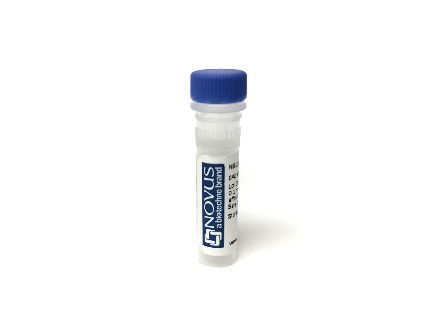
CD63 Antibody (SPM524) [Alexa Fluor® 405]

Novus Biologicals, part of Bio-Techne | Catalog # NBP2-34779AF405

Novus Biologicals, part of Bio-Techne
Catalog #
Availability
Size / Price
Qty
Loading...
NBP2-34779AF405

Key Product Details

Species Reactivity

Validated:

Human, Mouse

Applications

Flow Cytometry, Immunocytochemistry/ Immunofluorescence, Immunohistochemistry, Immunohistochemistry-Paraffin, Western Blot

Label

Alexa Fluor 405 (Excitation = 405 nm, Emission = 421 nm)

Antibody Source

Monoclonal Mouse IgG1 kappa Clone # SPM524

Concentration

Please see the vial label for concentration. If unlisted please contact technical services.

Product Summary for CD63 Antibody (SPM524) [Alexa Fluor® 405]

Immunogen

Smooth plasma membrane fraction of MeWo cells

Reactivity Notes

Mouse reactivity reported in scientific literature (PMID: 31207235).

Specificity

This monoclonal antibody recognizes protein of 26kDa-60kDa, which is identified as CD63. Its epitope is different from that of monoclonal antibody LAMP3/529. The tetraspanins are integral membrane proteins expressed on cell surface and granular membranes of hematopoietic cells and are components of multi-molecular complexes with specific integrins. The tetraspanin CD63 is a lysosomal membrane glycoprotein that translocates to the plasma membrane after platelet activation. CD63 is expressed on activated platelets, monocytes and macrophages, and is weakly expressed on granulocytes, T cell and B cells. It is located on the basophilic granule membranes and on the plasma membranes of lymphocytes and granulocytes. CD63 is a member of the TM4 superfamily of leukocyte glycoproteins that includes CD9, CD37 and CD53, which contain four transmembrane regions. CD63 may play a role in phagocytic and intracellular lysosome-phagosome fusion events. CD63 deficiency is associated with Hermansky-Pudlak syndrome and is strongly expressed during the early stages of melanoma progression.

Marker

Late Endosomes Marker

Clonality

Monoclonal

Host

Mouse

Isotype

IgG1 kappa

Applications for CD63 Antibody (SPM524) [Alexa Fluor® 405]

Application
Recommended Usage

Flow Cytometry

Optimal dilutions of this antibody should be experimentally determined.

Immunocytochemistry/ Immunofluorescence

Optimal dilutions of this antibody should be experimentally determined.

Immunohistochemistry

Optimal dilutions of this antibody should be experimentally determined.

Immunohistochemistry-Paraffin

Optimal dilutions of this antibody should be experimentally determined.

Western Blot

Optimal dilutions of this antibody should be experimentally determined.
Please Note: Optimal dilutions of this antibody should be experimentally determined.

Formulation, Preparation, and Storage

Purification

Protein A or G purified

Formulation

50mM Sodium Borate

Preservative

0.05% Sodium Azide

Concentration

Please see the vial label for concentration. If unlisted please contact technical services.

Shipping

The product is shipped with polar packs. Upon receipt, store it immediately at the temperature recommended below.

Stability & Storage

Store at 4C in the dark.

Background: CD63

CD63 (cluster of differentiation 63), also known as lysosome associated membrane protein 3 (LAMP-3), is a membrane spanning glycoprotein with a molecular mass ranging from 30-60 kDa that was the first characterized member of the tetraspanin superfamily (1,2). CD63 is ubiquitously expressed and largely present on the cell surface in the endosomal system (1-3). More specifically, it is generally present in multivesicular bodies (MVBs), also called late endosomes, and lysosomes (1). The CD63 molecule is a total of 238 amino acids (aa), has four hydrophobic membrane-spanning regions and three N-glycosylation sites, and the encoded protein has a theoretical molecular weight of 25 kDa (2,3). CD63 both directly and indirectly interacts with other proteins including integrins, cell surface receptors, other tetraspanins, kinases, and adapter proteins, to name a few (1). CD63 is involved in many cell processes including cell survival and activation, cell adhesion, invasion, and migration (1). Additionally, CD63 has been shown to interact with tissue inhibitor of metalloproteinase 1 (TIMP-1), which originally thought to be an inhibitor of cancer progression has recently been shown to have cancer promoting properties as well (1,4). The CD63-TIMP-1 interaction has been shown to activate the PI3K/AKT pathway in lung adenocarcinoma cells and also promote survival and invasion of acute myeloid leukemia cells (1).CD63 is a useful flow cytometry marker for basophil granulocytes and can be used for the basophil activation test (BAT) to assess IgE-mediated allergy response (5). As CD63 was initially discovered on activated blood platelets and is a lysosomal-associated protein it makes sense it would be involved in platelet or lysosomal-related disorders (1,2). More precisely, CD63 is associated with Hermansky-Pudlak syndrome, a disease characterized by albinism and platelet storage pool deficiency (6).

References

1. Pols, M. S., & Klumperman, J. (2009). Trafficking and function of the tetraspanin CD63. Experimental cell research. https://doi.org/10.1016/j.yexcr.2008.09.020

2. Metzelaar, M. J., Wijngaard, P. L., Peters, P. J., Sixma, J. J., Nieuwenhuis, H. K., & Clevers, H. C. (1991). CD63 antigen. A novel lysosomal membrane glycoprotein, cloned by a screening procedure for intracellular antigens in eukaryotic cells. The Journal of biological chemistry.

3. Horejsi, V., & Vlcek, C. (1991). Novel structurally distinct family of leucocyte surface glycoproteins including CD9, CD37, CD53 and CD63. FEBS letters. https://doi.org/10.1016/0014-5793(91)80988-f

4. Eckfeld, C., HauBler, D., Schoeps, B., Hermann, C. D., & Kruger, A. (2019). Functional disparities within the TIMP family in cancer: hints from molecular divergence. Cancer metastasis reviews. https://doi.org/10.1007/s10555-019-09812-6

5. Hoffmann, H. J., Santos, A. F., Mayorga, C., Nopp, A., Eberlein, B., Ferrer, M., Rouzaire, P., Ebo, D. G., Sabato, V., Sanz, M. L., Pecaric-Petkovic, T., Patil, S. U., Hausmann, O. V., Shreffler, W. G., Korosec, P., & Knol, E. F. (2015). The clinical utility of basophil activation testing in diagnosis and monitoring of allergic disease. Allergy. https://doi.org/10.1111/all.12698

6. Dell'Angelica, E. C., Shotelersuk, V., Aguilar, R. C., Gahl, W. A., & Bonifacino, J. S. (1999). Altered trafficking of lysosomal proteins in Hermansky-Pudlak syndrome due to mutations in the beta 3A subunit of the AP-3 adaptor. Molecular cell. https://doi.org/10.1016/s1097-2765(00)80170-7

Alternate Names

CD63, Granulophysin, Lamp-3, ME491, OMA81H, Tspan30

Gene Symbol

CD63

Additional CD63 Products

Product Documents for CD63 Antibody (SPM524) [Alexa Fluor® 405]

Certificate of Analysis

To download a Certificate of Analysis, please enter a lot number in the search box below.

Product Specific Notices for CD63 Antibody (SPM524) [Alexa Fluor® 405]

Alexa Fluor (R) products are provided under an intellectual property license from Life Technologies Corporation. The purchase of this product conveys to the buyer the non-transferable right to use the purchased product and components of the product only in research conducted by the buyer (whether the buyer is an academic or for-profit entity). The sale of this product is expressly conditioned on the buyer not using the product or its components, or any materials made using the product or its components, in any activity to generate revenue, which may include, but is not limited to use of the product or its components: (i) in manufacturing; (ii) to provide a service, information, or data in return for payment; (iii) for therapeutic, diagnostic or prophylactic purposes; or (iv) for resale, regardless of whether they are resold for use in research. For information on purchasing a license to this product for purposes other than as described above, contact Life Technologies Corporation, 5791 Van Allen Way, Carlsbad, CA 92008 USA or outlicensing@lifetech.com. This conjugate is made on demand. Actual recovery may vary from the stated volume of this product. The volume will be greater than or equal to the unit size stated on the datasheet.

This product is for research use only and is not approved for use in humans or in clinical diagnosis. Primary Antibodies are guaranteed for 1 year from date of receipt.

Loading...
Loading...
Loading...
Loading...
Loading...
Loading...