CD68/SR-D1 Antibody (KP1) - BSA Free
Novus Biologicals, part of Bio-Techne | Catalog # NB100-683
Clone KP1 was used by HLDA to establish CD designation.

![Immunohistochemistry: CD68/SR-D1 Antibody (KP1) - BSA Free [NB100-683] Immunohistochemistry: CD68/SR-D1 Antibody (KP1) - BSA Free [NB100-683]](https://resources.bio-techne.com/images/products/CD68-SR-D1-Antibody-KP1-Immunohistochemistry-NB100-683-img0009.jpg)
Conjugate
Catalog #
Key Product Details
Species Reactivity
Validated:
Human, Mouse, Rat
Cited:
Human, Mouse, Rat
Applications
Validated:
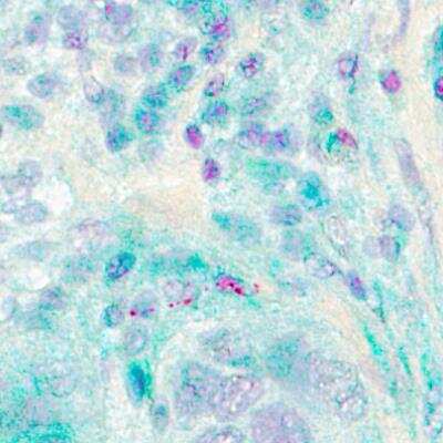
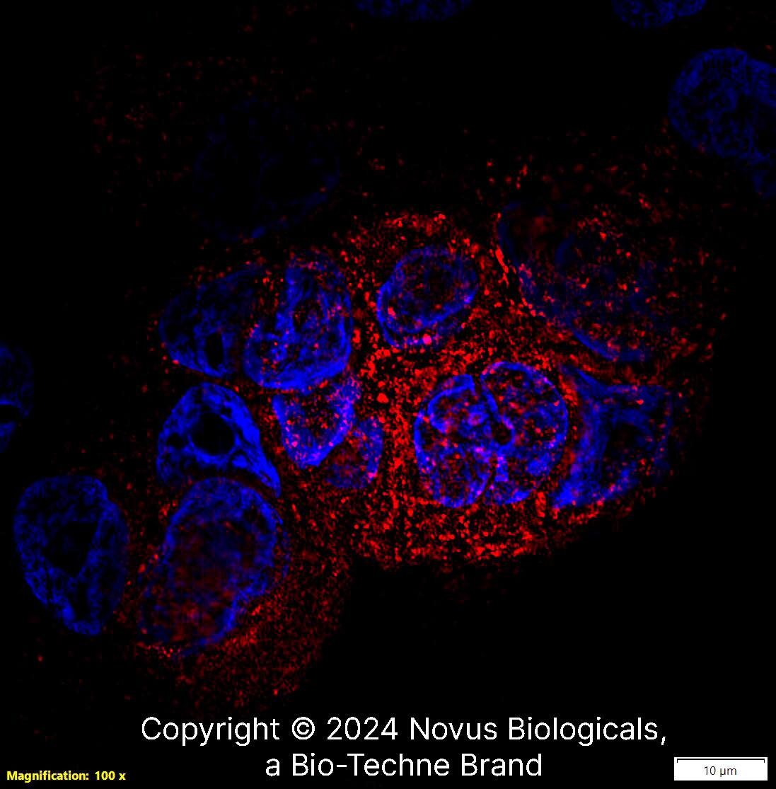
Dual RNAscope ISH-IHC, Electron Microscopy, Flow Cytometry, Immunocytochemistry/ Immunofluorescence, Immunohistochemistry, Immunohistochemistry-Frozen, Immunohistochemistry-Paraffin, Immunoprecipitation, Western Blot
Cited:
Flow Cytometry, IF/IHC, Immunocytochemistry/ Immunofluorescence, Immunofluorescence, Immunohistochemistry, Immunohistochemistry-Frozen, Immunohistochemistry-Paraffin, In vivo assay, Western Blot
Label
Unconjugated
Antibody Source
Monoclonal Mouse IgG1 kappa Clone # KP1
Format
BSA Free
Concentration
1.0 mg/ml
Product Specifications
Immunogen
This CD68/SR-D1 Antibody (KP1) was developed against subcellular fraction of human alveolar macrophages.
Reactivity Notes
Rat reactivity reported in scientific literature (PMID: 25058444)
Localization
Cell membrane
Specificity
This CD68/SR-D1 Antibody (KP1) is specific to macrophages in a wide variety of human tissues. It reacts with myeloid precursors and peripheral blood granulocytes. It also stains a cell population known as Plasmacytoid T cells.
Marker
Macrophage Marker
Clonality
Monoclonal
Host
Mouse
Isotype
IgG1 kappa
Scientific Data Images for CD68/SR-D1 Antibody (KP1) - BSA Free
Immunohistochemistry: CD68/SR-D1 Antibody (KP1) - BSA Free [NB100-683]
Immunohistochemistry: CD68/SR-D1 Antibody (KP1) [NB100-683] - Representative images (40x) of infiltrated macrophages stained with anti-CD 68 in the gingiva. The black arrowheads indicate the infiltrated macrophages. The right graphs show the quantitative data of CD68-positive cells in HPF (magnification of 200x). Data are expressed as the mean +/- SEM of three slides. *p<0.05 compared with control cells. Image collected and cropped by CiteAb from the following publication (//doi.org/10.1371/journal.pone.0102450) licensed under a CC-BY license.Flow Cytometry: CD68/SR-D1 Antibody (KP1) - BSA Free [NB100-683]
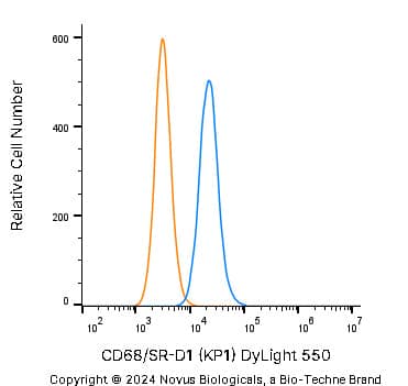
Flow Cytometry: CD68/SR-D1 Antibody (KP1) [NB100-683] - An intracellular stain was performed on THP-1 cells with CD68/SR-D1 Antibody NB100-683 (blue) and a matched isotype control (orange). Cells were fixed with 4% PFA and then permeabilized with 0.1% saponin. Cells were incubated in an antibody dilution of 1:50 for 30 minutes at room temperature, followed by Mouse IgG (H+L) Cross-Adsorbed Secondary Antibody.Immunocytochemistry/ Immunofluorescence: CD68/SR-D1 Antibody (KP1) - BSA Free [NB100-683]
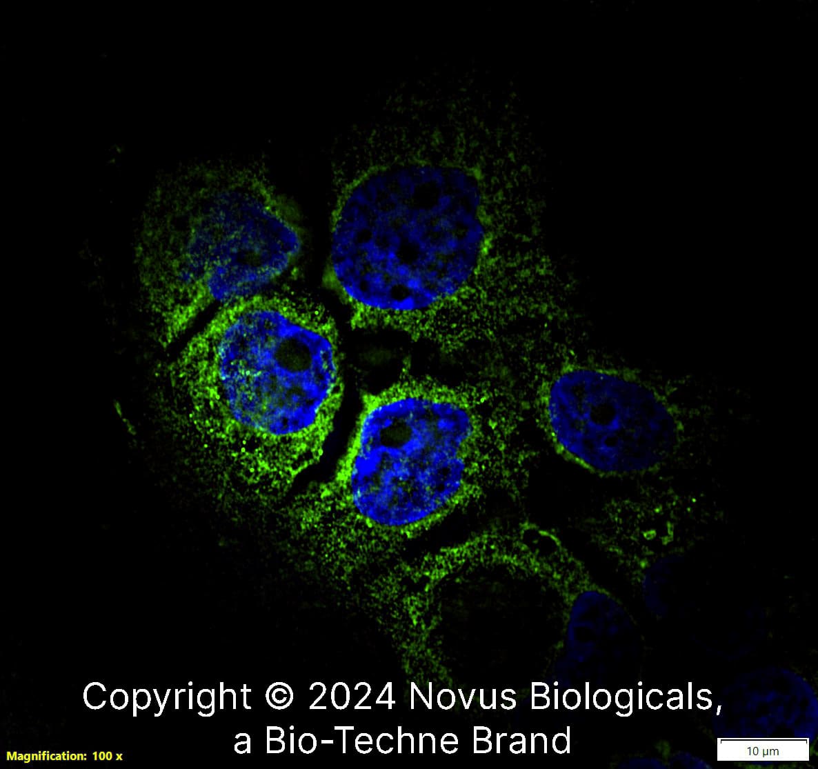
Immunocytochemistry/Immunofluorescence: CD68/SR-D1 Antibody (KP1) [NB100-683] - A431 cells were fixed in 4% paraformaldehyde for 10 minutes and permeabilized in 0.05% Triton X-100 in PBS for 5 minutes. The cells were incubated with CD68/SR-D1 Antibody [KP1] (NB100-683) at 1 ug/ml overnight at 4C and detected with an anti-mouse Dylight 488 (Green) at a 1:1000 dilution for 60 minutes. Nuclei were counterstained with DAPI (Blue). Cells were imaged using a 100X objective and digitally deconvolved.Applications for CD68/SR-D1 Antibody (KP1) - BSA Free
Application
Recommended Usage
Electron Microscopy
reported in scientific literature (PMID 8962141)
Flow Cytometry
reported in scientific literature (PMID 18405323)
Immunocytochemistry/ Immunofluorescence
1-5 ug/ml
Immunohistochemistry
1:100-1:200
Immunohistochemistry-Frozen
1:100-1:200
Immunohistochemistry-Paraffin
1:100-1:200
Western Blot
2 ug/ml
Application Notes
IHC: For an ABC system, dilute 1:20 and incubate for 30-60 minutes at RT. IHC-P tissue sections require high temperature antigen unmasking with 10 mM citrate buffer, pH 6.0 prior to immunostaining.
Reviewed Applications
Read 1 review rated 5 using NB100-683 in the following applications:
Formulation, Preparation, and Storage
Purification
Protein G purified
Formulation
PBS
Format
BSA Free
Preservative
0.02% Sodium Azide
Concentration
1.0 mg/ml
Shipping
The product is shipped with polar packs. Upon receipt, store it immediately at the temperature recommended below.
Stability & Storage
Store at 4C. Do not freeze.
Background: CD68/SR-D1
CD68 is highly expressed in cells of the mononuclear phagocyte system such as macrophages, microglia, osteoclasts, and myeloid dendritic cells (DCs); and is expressed to a lesser extent in lymphoid cells (CD19+ B lymphocytes and CD4+ T lymphocytes), human umbilical cord mesenchymal stem cells (MSCs), fibroblasts, endothelial cells, multiple non-hematopoietic cancer cell lines, and human arterial intimal smooth muscle cells (SMCs). Expression has been also observed in diseased states for granulocytes and neutrophils, in particular basophils from myeloproliferative disorders and intestinal neutrophils from inflammatory bowel disease (IBD), respectively (1).
Although the function of CD68 has yet to be established, it has often been used as an immunohistochemistry (IHC) marker of inflammation and for granular cell tumors (GCTs). CD68+ tumor associated macrophages (TAMs) has been suggested to be a predictive marker for poor cancer prognosis, but a meta-analysis showed the presence of CD68 is not correlated with survival (2). In addition, a role in hepatic malaria infection has been reported based on the finding that peptide P39 binds CD68, considered a receptor for malaria sporozoite, and inhibits parasite entry into Kupffer cells. CD68 was deemed a member of the Scavenger receptor family due to its upregulation in macrophages following inflammatory stimuli, ability to bind modified LDL, phosphatidylserine, and apoptotic cells, as well as shuttling between the plasma membrane and endosomes. CD68 has been linked to atherogenesis based on binding and internalization of its ligand, oxLDL (1).
References
1. Chistiakov, DA, Killingsworth, MC, Myasoedova, VA. Orekhov AN, Bobryshev YV. (2017) CD68/macrosialin: not just a histochemical marker. Lab Invest. 97:4-13. PMID: 27869795
2. Troiano G, Caponio VCA, Adipietro I, Tepedino M, Santoro R, Laino L, Lo Russo L, Cirillo N, Lo Muzio L. (2019) Prognostic significance of CD68+ and CD163+ tumor associated macrophages in head and neck squamous cell carcinoma: A systematic review and meta-analysis. Oral Oncol. 93:66-75. PMID: 31109698.
Alternate Names
CD68, gp110, Macrosialin, SCARD1, SR-D1, SRD1
Entrez Gene IDs
968 (Human)
Gene Symbol
CD68
UniProt
Additional CD68/SR-D1 Products
Product Documents for CD68/SR-D1 Antibody (KP1) - BSA Free
Product Specific Notices for CD68/SR-D1 Antibody (KP1) - BSA Free
This product is for research use only and is not approved for use in humans or in clinical diagnosis. Primary Antibodies are guaranteed for 1 year from date of receipt.
Loading...
Loading...
Loading...
Loading...
Loading...
![Flow Cytometry: CD68/SR-D1 Antibody (KP1) - BSA Free [NB100-683] Flow Cytometry: CD68/SR-D1 Antibody (KP1) - BSA Free [NB100-683]](https://resources.bio-techne.com/images/products/CD68-SR-D1-Antibody-KP1-Flow-Cytometry-NB100-683-img0004.jpg)
![Immunocytochemistry/ Immunofluorescence: CD68/SR-D1 Antibody (KP1) - BSA Free [NB100-683] Immunocytochemistry/ Immunofluorescence: CD68/SR-D1 Antibody (KP1) - BSA Free [NB100-683]](https://resources.bio-techne.com/images/products/CD68-SR-D1-Antibody-KP1-Immunocytochemistry-Immunofluorescence-NB100-683-img0012.jpg)
![Western Blot: CD68/SR-D1 Antibody (KP1)BSA Free [NB100-683] Western Blot: CD68/SR-D1 Antibody (KP1)BSA Free [NB100-683]](https://resources.bio-techne.com/images/products/CD68-SR-D1-Antibody-KP1-Western-Blot-NB100-683-img0007.jpg)
![Flow Cytometry: CD68/SR-D1 Antibody (KP1) - BSA Free [NB100-683] Flow Cytometry: CD68/SR-D1 Antibody (KP1) - BSA Free [NB100-683]](https://resources.bio-techne.com/images/products/CD68-SR-D1-Antibody-KP1-Flow-Cytometry-NB100-683-img0013.jpg)
![Immunocytochemistry/ Immunofluorescence: CD68/SR-D1 Antibody (KP1) - BSA Free [NB100-683] Immunocytochemistry/ Immunofluorescence: CD68/SR-D1 Antibody (KP1) - BSA Free [NB100-683]](https://resources.bio-techne.com/images/products/CD68-SR-D1-Antibody-KP1-Immunocytochemistry-Immunofluorescence-NB100-683-img0011.jpg)
![Immunohistochemistry-Paraffin: CD68/SR-D1 Antibody (KP1) - BSA Free [NB100-683] Immunohistochemistry-Paraffin: CD68/SR-D1 Antibody (KP1) - BSA Free [NB100-683]](https://resources.bio-techne.com/images/products/CD68-SR-D1-Antibody-KP1-Immunohistochemistry-Paraffin-NB100-683-img0006.jpg)
![Flow Cytometry: CD68/SR-D1 Antibody (KP1) - BSA Free [NB100-683] Flow Cytometry: CD68/SR-D1 Antibody (KP1) - BSA Free [NB100-683]](https://resources.bio-techne.com/images/products/CD68-SR-D1-Antibody-KP1-Flow-Cytometry-NB100-683-img0005.jpg)


