Cytokeratin 5 Antibody - BSA Free
Novus Biologicals, part of Bio-Techne | Catalog # NBP2-61931

![Western Blot: Cytokeratin 5 AntibodyBSA Free [NBP2-61931] Western Blot: Cytokeratin 5 AntibodyBSA Free [NBP2-61931]](https://resources.bio-techne.com/images/products/Cytokeratin-5-Antibody-Western-Blot-NBP2-61931-img0002.jpg)
Conjugate
Catalog #
Key Product Details
Species Reactivity
Validated:
Human, Rat
Cited:
Human
Applications
Validated:
Flow Cytometry, Immunocytochemistry/ Immunofluorescence, Immunohistochemistry, Immunohistochemistry-Paraffin, Western Blot
Cited:
Immunohistochemistry-Frozen
Label
Unconjugated
Antibody Source
Polyclonal Rabbit IgG
Format
BSA Free
Concentration
1.0 mg/ml
Product Specifications
Immunogen
Partial recombinant human Cytokeratin 5 protein (amino acids 19-78)
Clonality
Polyclonal
Host
Rabbit
Isotype
IgG
Scientific Data Images for Cytokeratin 5 Antibody - BSA Free
Western Blot: Cytokeratin 5 AntibodyBSA Free [NBP2-61931]
Western Blot: Cytokeratin 5 Antibody [NBP2-61931] - Total protein from human A431 cells and rat PC12 cells was separated on a 7.5% gel by SDS-PAGE, transferred to PVDF membrane and blocked in 5% non-fat milk in TBST. The membrane was probed with 2.0 ug/ml anti-Cytokeratin 5 in 5% non-fat milk in TBST and detected with an anti-rabbit HRP secondary antibody using chemiluminescence.Immunocytochemistry/ Immunofluorescence: Cytokeratin 5 Antibody - BSA Free [NBP2-61931]
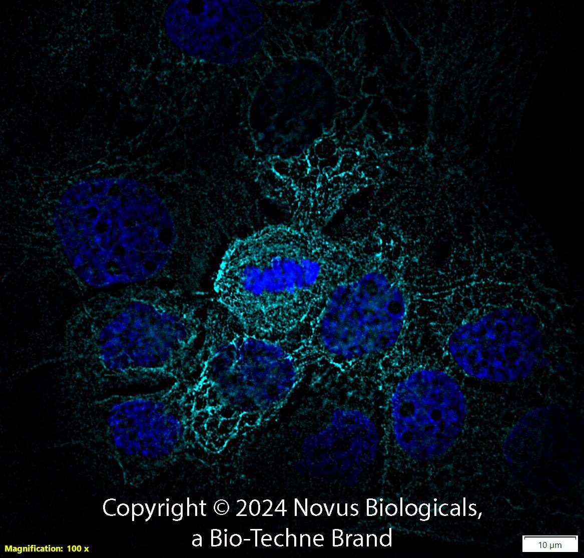
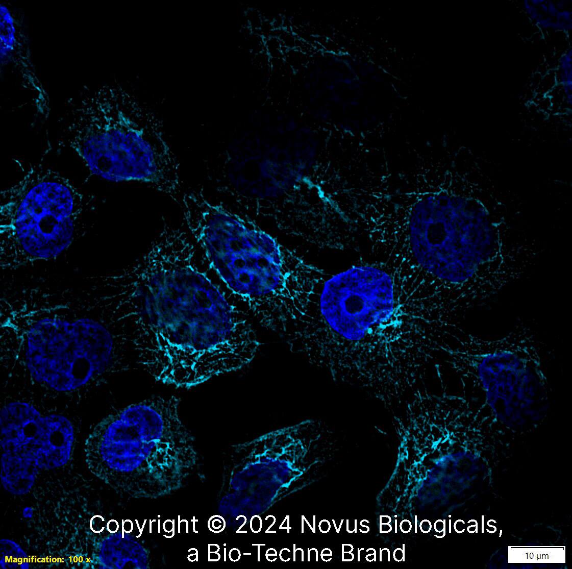
Immunocytochemistry/Immunofluorescence: Cytokeratin 5 Antibody [NBP2-61931] - A431 cells were fixed for 10 minutes using 10% formalin and then permeabilized for 5 minutes using 1X PBS + 0.1% Triton X-100. The cells were incubated with anti-Cytokeratin 5 at 5 ug/ml overnight at 4C and detected with an anti-rabbit DyLight 488 (Green) at a 1:500 dilution. Alpha tubulin (DM1A) NB100-690 was used as a co-stain at a 1:1000 dilution and detected with an anti-mouse DyLight 550 (Red) at a 1:500 dilution. Nuclei were counterstained with DAPI (Blue). Cells were imaged using a 40X objective.Immunohistochemistry-Paraffin: Cytokeratin 5 Antibody - BSA Free [NBP2-61931]
Immunohistochemistry-Paraffin: Cytokeratin 5 Antibody [NBP2-61931] - Analysis of a FFPE tissue section of human skin using 1:1000 dilution of rabbit anti-Cytokeratin 5 antibody. The staining was developed using HRP labeled anti-rabbit secondary antibody and DAB reagent, and nuclei of cells were counter-stained with hematoxylin. This Cytokeratin 5 antibody generated a specific membrane cytoplasmic staining in the epithelial cells of the skin tissue. The signal was highest in the keratinocytes while dermis was largely negative for this Cytokeratin 5 (KRT5).Applications for Cytokeratin 5 Antibody - BSA Free
Application
Recommended Usage
Flow Cytometry
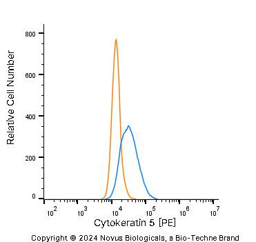
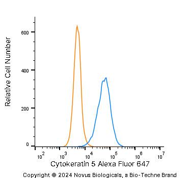
2-10 ug/million cells
Immunocytochemistry/ Immunofluorescence
5 ug/ml
Immunohistochemistry
1:1000
Immunohistochemistry-Paraffin
1:1000
Western Blot
1 ug/ml
Formulation, Preparation, and Storage
Purification
Immunogen affinity purified
Formulation
PBS
Format
BSA Free
Preservative
0.02% Sodium Azide
Concentration
1.0 mg/ml
Shipping
The product is shipped with polar packs. Upon receipt, store it immediately at the temperature recommended below.
Stability & Storage
Store at 4C short term. Aliquot and store at -20C long term. Avoid freeze-thaw cycles.
Background: Cytokeratin 5
Alternate Names
CK-5, DDD, EBS2, Keratin 5, KRT5
Gene Symbol
KRT5
Additional Cytokeratin 5 Products
Product Documents for Cytokeratin 5 Antibody - BSA Free
Product Specific Notices for Cytokeratin 5 Antibody - BSA Free
This product is for research use only and is not approved for use in humans or in clinical diagnosis. Primary Antibodies are guaranteed for 1 year from date of receipt.
Loading...
Loading...
Loading...
Loading...
Loading...
![Immunocytochemistry/ Immunofluorescence: Cytokeratin 5 Antibody - BSA Free [NBP2-61931] Immunocytochemistry/ Immunofluorescence: Cytokeratin 5 Antibody - BSA Free [NBP2-61931]](https://resources.bio-techne.com/images/products/Cytokeratin-5-Antibody-Immunocytochemistry-Immunofluorescence-NBP2-61931-img0001.jpg)
![Immunohistochemistry-Paraffin: Cytokeratin 5 Antibody - BSA Free [NBP2-61931] Immunohistochemistry-Paraffin: Cytokeratin 5 Antibody - BSA Free [NBP2-61931]](https://resources.bio-techne.com/images/products/Cytokeratin-5-Antibody-Immunohistochemistry-Paraffin-NBP2-61931-img0003.jpg)
![Flow Cytometry: Cytokeratin 5 Antibody - BSA Free [NBP2-61931] Flow Cytometry: Cytokeratin 5 Antibody - BSA Free [NBP2-61931]](https://resources.bio-techne.com/images/products/Cytokeratin-5-Antibody-Flow-Cytometry-NBP2-61931-img0004.jpg)


