EpCAM/TROP1 Antibody (EGP40/826) - BSA Free
Novus Biologicals, part of Bio-Techne | Catalog # NBP2-44643

![Immunohistochemistry-Paraffin: EpCAM/TROP1 Antibody (EGP40/826) - BSA Free [NBP2-44643] Immunohistochemistry-Paraffin: EpCAM/TROP1 Antibody (EGP40/826) - BSA Free [NBP2-44643]](https://resources.bio-techne.com/images/products/EpCAM-TROP1-Antibody-EGP40-826-Immunohistochemistry-Paraffin-NBP2-44643-img0003.jpg)
Conjugate
Catalog #
Forumulation
Catalog #
Key Product Details
Species Reactivity
Human, Mouse (Negative), Rat (Negative)
Applications
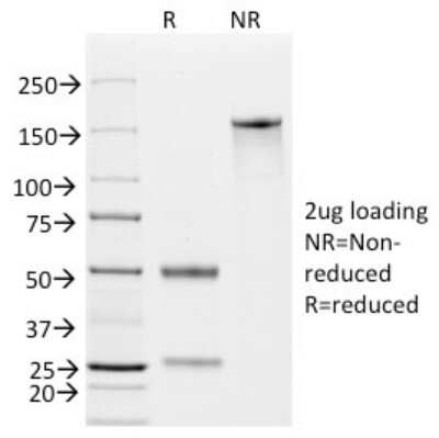
Flow Cytometry, Immunocytochemistry/ Immunofluorescence, Immunohistochemistry, Immunohistochemistry-Paraffin, SDS-Page, Western Blot
Label
Unconjugated
Antibody Source
Monoclonal Mouse IgG1 kappa Clone # EGP40/826
Format
BSA Free
Concentration
1.0 mg/ml
Product Specifications
Immunogen
A synthetic peptide (around aa 20-60) from the N-terminus of human TACSTD1 protein.
Localization
Cell surface
Specificity
Recognizes a 40-43kDa transmembrane epithelial glycoprotein, identified as epithelial specific antigen (ESA), or epithelial cellular adhesion molecule (Ep-CAM). Ep-CAM is expressed on baso-lateral cell surface in most simple epithelia and a vast majority of carcinomas. This antibody has been used to distinguish adenocarcinoma from pleural mesothelioma and hepatocellular carcinoma. It is also useful in distinguishing serous carcinomas of the ovary from mesothelioma. This epithelial antigen plays an important role as a tumor-cell marker in lymph nodes from patients with esophageal carcinoma otherwise classified as node-negative. Epithelial antigen has also been suggested as a discriminator between basal cell and baso-squamous carcinomas, and squamous cell carcinoma of the skin.
Marker
Epithelial Marker
Clonality
Monoclonal
Host
Mouse
Isotype
IgG1 kappa
Theoretical MW
41 kDa.
Disclaimer note: The observed molecular weight of the protein may vary from the listed predicted molecular weight due to post translational modifications, post translation cleavages, relative charges, and other experimental factors.
Disclaimer note: The observed molecular weight of the protein may vary from the listed predicted molecular weight due to post translational modifications, post translation cleavages, relative charges, and other experimental factors.
Scientific Data Images for EpCAM/TROP1 Antibody (EGP40/826) - BSA Free
Immunohistochemistry-Paraffin: EpCAM/TROP1 Antibody (EGP40/826) - BSA Free [NBP2-44643]
Immunohistochemistry-Paraffin: EpCAM/TROP1 Antibody (EGP40/826) [NBP2-44643] - Human Breast Carcinoma stained with Ep-CAM Monoclonal Antibody (EGP40/826).Immunofluorescence: EpCAM/TROP1 Antibody (EGP40/826) - BSA Free [NBP2-44643]
Immunofluorescence: EpCAM/TROP1 Antibody (EGP40/826) [NBP2-44643] - Confocal Immunofluorescent analysis of SK-OV-3 cells using AF488-labeled EpCAM Monoclonal Antibody (EGP40/826) (Green). F-actin filaments were labeled with DyLight 554 Phalloidin (red). DAPI was used to stain the cell nuclei (blue).Immunofluorescence: EpCAM/TROP1 Antibody (EGP40/826) - BSA Free [NBP2-44643]
Immunofluorescence: EpCAM/TROP1 Antibody (EGP40/826) [NBP2-44643] - Confocal Immunofluorescent analysis of SK-OV-3 cells using AF488-labeled Isotype Control Monoclonal Antibody (IgG1) (Green). DAPI was used to stain the cell nuclei (blue). (Negative Control)Applications for EpCAM/TROP1 Antibody (EGP40/826) - BSA Free
Application
Recommended Usage
Flow Cytometry
0.5 - 1 ug/million cells in 0.1 ml
Immunohistochemistry-Paraffin
0.5 - 1.0 ug/ml
Western Blot
0.5 - 1.0 ug/ml
Application Notes
Hu-chromosome location: 2q21
Molecular weight of antigen: 40-43kDa
Immunohistochemistry-Paraffin 0.5 - 1 ug/ml for 30 minutes at RT; Staining of formalin-fixed tissues requires boiling tissue sections in 10mM Citrate Buffer, pH 6.0, for 10 - 20 min followed by cooling at RT for 20 minutes. The 7mL size is a pre-diluted size and no additional dilutions are required before using this item for the intended application.
Formulation, Preparation, and Storage
Purification
Protein A or G purified
Formulation
PBS
Format
BSA Free
Preservative
0.02% Sodium Azide
Concentration
1.0 mg/ml
Shipping
The product is shipped with polar packs. Upon receipt, store it immediately at the temperature recommended below.
Stability & Storage
Store at 4C short term. Aliquot and store at -20C long term. Avoid freeze-thaw cycles.
Background: EpCAM/TROP1
EpCAM functions as an intracellular signaling molecule and contributes to regulation of epithelial-to-mesenchymal (EMT) transition (4,5). EpCAM is cleaved during the process of regulated intramembrane proteolysis (RIP) (4,5). Initial cleavage occurs at the membrane via a disintegrin and metalloprotease 17 (ADAM17) which releases EpCAM's extracellular domain (EpEx) (4,5). A secondary cleavage is mediated by presenilin 2 (PSEN2) which releases EpCAM's cytoplasmic trail (EpICD) (4,5). EpICD translocates to the nucleus where it associates with beta-catenin, FHL-2, and LEF-1 and induces transcription of genes related to EMT and tumor growth (4,5). EpCAM has also been shown to regulate structure and functionality of the apical junction complex of cells through direct interaction with claudin-7 and association with E-cadherin (2,5). Loss of EpCAM disrupts adherins junction and tight junction structure and function (2).
References
1. Mohtar MA, Syafruddin SE, Nasir SN, Low TY. Revisiting the Roles of Pro-Metastatic EpCAM in Cancer. Biomolecules. 2020; 10(2):255. https://doi.org/10.3390/biom10020255
2. Huang L, Yang Y, Yang F, et al. Functions of EpCAM in physiological processes and diseases (Review). Int J Mol Med. 2018; 42(4):1771-1785. https://doi.org/10.3892/ijmm.2018.3764
3. Brown TC, Sankpal NV, Gillanders WE. Functional Implications of the Dynamic Regulation of EpCAM during Epithelial-to-Mesenchymal Transition. Biomolecules. 2021; 11(7):956. https://doi.org/10.3390/biom11070956
4. Uniprot (P16422)
5. Schnell U, Cirulli V, Giepmans BN. EpCAM: structure and function in health and disease. Biochim Biophys Acta. 2013; 1828(8):1989-2001. https://doi.org/10.1016/j.bbamem.2013.04.018
Long Name
Epithelial Cell Adhesion Molecule
Alternate Names
17-1A, CD326, GA733-2, gp40, KS1/4, M4S1, TACSTD1, TROP1
Gene Symbol
EPCAM
UniProt
Additional EpCAM/TROP1 Products
Product Documents for EpCAM/TROP1 Antibody (EGP40/826) - BSA Free
Product Specific Notices for EpCAM/TROP1 Antibody (EGP40/826) - BSA Free
This product is for research use only and is not approved for use in humans or in clinical diagnosis. Primary Antibodies are guaranteed for 1 year from date of receipt.
Loading...
Loading...
Loading...
Loading...
Loading...
![Immunofluorescence: EpCAM/TROP1 Antibody (EGP40/826) - BSA Free [NBP2-44643] Immunofluorescence: EpCAM/TROP1 Antibody (EGP40/826) - BSA Free [NBP2-44643]](https://resources.bio-techne.com/images/products/EpCAM-TROP1-Antibody-EGP40-826-Immunofluorescence-NBP2-44643-img0001.jpg)
![Immunofluorescence: EpCAM/TROP1 Antibody (EGP40/826) - BSA Free [NBP2-44643] Immunofluorescence: EpCAM/TROP1 Antibody (EGP40/826) - BSA Free [NBP2-44643]](https://resources.bio-techne.com/images/products/EpCAM-TROP1-Antibody-EGP40-826-Immunofluorescence-NBP2-44643-img0002.jpg)
