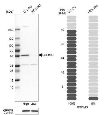
Western Blot: GSDMDC1 Antibody [NBP2-33422] -
Western Blot: GSDMDC1 Antibody [NBP2-33422] - miR-513c-5p negatively regulates caspase-1 expression in HUVECs. (A) The expression of miR-513c-5p after NC, miR-513c-5p mimics, INC & miR-513c-5p inhibitor transfection was evaluated by qRT-PCR. (B) The expression level of caspase-1 mRNA after NC, miR-513c-5p mimics, INC & miR-513c-5p inhibitor transfection was detected by qRT-PCR. (C,D) Expression of caspase-1 in HUVECs were detected after transfection NC, miR-513c-5p mimics, INC, miR-513c-5p inhibitor by immunofluorescence staining (magnification, ×40). Scale bar = 200 μm. (E,F) Protein expression levels of caspase-1 & GSDMD were examined by Western blot. (G) The expression of IL-1 beta & IL-18 was detected by ELISA. *p < 0.05, **p < 0.01, & ***p < 0.001, ****p < 0.0001. Image collected & cropped by CiteAb from the following publication (https://pubmed.ncbi.nlm.nih.gov/35445025), licensed under a CC-BY license. Not internally tested by Novus Biologicals.
Western Blot: GSDMDC1 Antibody [NBP2-33422] -
Western Blot: GSDMDC1 Antibody [NBP2-33422] - Pyroptosome formation in the cortex & hippocampus of aged mice: Aging induces laddering of ASC in cortex (a) & hippocampus (b) of mice, indicating formation of the pyroptosome, an oligomerization of ASC that leads to pyroptosis. Representative immunoblot & quantification of gasdermin-D in the cortex (c) & hippocampus (d) of aged mice when compared to young. Gasdermin-D is significantly elevated in the cortex & hippocampus of aged mice. Data presented as mean+/-SEM. N = 5 per group. * p < 0.05 Image collected & cropped by CiteAb from the following publication (https://pubmed.ncbi.nlm.nih.gov/30473634), licensed under a CC-BY license. Not internally tested by Novus Biologicals.
Western Blot: GSDMDC1 Antibody [NBP2-33422] -
Western Blot: GSDMDC1 Antibody [NBP2-33422] - Expressions of caspase-1, GSDMD, IL-1 beta, & IL-18 are up-regulated in DVT patients. (A) mRNA level of caspase-1 in the PBMCs from 30 DVT patients & 30 controls was determined by qRT-PCR. (B) Caspase-1 & GSDMD protein levels were measured in PBMCs of DVT subjects & controls by Western blot. (C,D) Protein levels of IL-1 beta & IL-18 in the serum from 12 DVT patients & 12 controls were determined by ELISA. ****p < 0.0001. Image collected & cropped by CiteAb from the following publication (https://pubmed.ncbi.nlm.nih.gov/35445025), licensed under a CC-BY license. Not internally tested by Novus Biologicals.
Western Blot: GSDMDC1 Antibody [NBP2-33422] -
Western Blot: GSDMDC1 Antibody [NBP2-33422] - Overexpression of miR-513c-5p suppresses pyroptosis of VECs & DVT formation by inhibiting caspase-1. (A) Expression of miR-513c-5p in vascular tissue was detected by qRT-PCR in DVT mice treated with NC, miR-513c-5p mimics, respectively. (B) Relative expression of miR-513c-5p was measured by qRT-PCR in PBMCs of DVT each treatment group. (C) Confocal microscopy images of miR-513c-5p expression in vascular tissues (miR-513c-5p, red; DAPI, blue) (magnification, ×200). Scale bars = 100 μm. (D) H&E staining of serial cross sections of inferior vena cava (IVC) from DVT mice treated with NC, miR-513c-5p mimics at 48 h (magnification, ×40), respectively. Scale bars = 500 μm. (E) Representative images of thrombi in each treatment group were detected by vascular ultrasound at 48 h post-operation. (F) Thrombus length & weight at 48 h post-operation in DVT mice (n = 15) treated with NC, miR-513c-5p mimics. (G) Caspase-1 mRNA level was determined by qRT-PCR in vascular tissue of each treatment group. (H) Caspase-1 & GSDMD protein levels were determined by Western blot in vascular tissue of DVT each treatment group. (I) IL-1 beta & IL-18 protein levels in vascular tissue were determined by ELISA in DVT each treatment group. **p < 0.01, ***p < 0.001, & ****p < 0.0001. Image collected & cropped by CiteAb from the following publication (https://pubmed.ncbi.nlm.nih.gov/35445025), licensed under a CC-BY license. Not internally tested by Novus Biologicals.
Western Blot: GSDMDC1 Antibody [NBP2-33422] -
Western Blot: GSDMDC1 Antibody [NBP2-33422] - Caspase-1 inhibitor (vx-765) inhibits the formation of DVT in vivo. (A,B) H&E staining of serial cross sections of IVC from Ctrl, Sham, DVT, DVT+vehicle, DVT+vx-765, DVT+vehicle+miR-513c-5p inhibitor, DVT+vx-765+miR-513c-5p inhibitor at 48 h (magnification, ×40). Scale bars = 500 μm. (C,D) Thrombus length & weight at 48 h post-operation in the different treatment groups after the administration of caspase-1 inhibitor (vx-765) (n = 15 in each group). (E) Representative images of thrombi in each treatment group were detected by vascular ultrasound at 48 h post-operation. (F) Caspase-1 & GSDMD protein levels were determined by Western blot in vascular tissue of DVT animal group & vx-765-treated groups. (G) Expression of IL-1 beta & IL-18 in vascular tissue were detected by ELISA with vx-765 treated each group. **p < 0.01, ***p < 0.001, ****p < 0.0001. Image collected & cropped by CiteAb from the following publication (https://pubmed.ncbi.nlm.nih.gov/35445025), licensed under a CC-BY license. Not internally tested by Novus Biologicals.
Western Blot: GSDMDC1 Antibody [NBP2-33422] -
Western Blot: GSDMDC1 Antibody [NBP2-33422] - mIL-18 release from unprimed monocytes is dependent on GSDMD. (A, B) Unprimed THP-1 cells pre-incubated with punicalagin (50 µM, 15 min) prior to treatment with nigericin (10 µM, 45 min). (C, D) Unprimed WT & GSDMD KO THP-1s were treated with nigericin (10 µM, 45 min). (A, C) Secreted IL-18 was measured by ELISA & cell death was measured by LDH assay & shown as percentage relative to total cell death, n=3 independent biological replicates, mean ± S.D., *P < 0.05; ***P <0.001; ****P <0.0001; ns (not significant) using one-way ANOVA comparing each sample to nigericin only treated sample (A) or two-way ANOVA comparing UT to nigericin treated WT THP-1s as well as nigericin treated WT THP-1s to nigericin treated GSDMD KO THP-1s. (C). (B, D) Western blot analysis of THP-1 cells for mIL-18 (18 kDa), pro-IL-18 (24 kDa), mCaspase-1 (20 kDa), pro-Caspase-1 (45 kDa), GSDMD full length (FL, 53 kDa), GSDMD N-terminus (NT, 31 kDa), as well as loading control beta-actin (42 kDa). Blots are representative of at least 3 independent experiments. Image collected & cropped by CiteAb from the following publication (https://pubmed.ncbi.nlm.nih.gov/33101286), licensed under a CC-BY license. Not internally tested by Novus Biologicals.
Western Blot: GSDMDC1 Antibody [NBP2-33422] -
Western Blot: GSDMDC1 Antibody [NBP2-33422] - Priming is not required for NLRP3 inflammasome activation in human monocytes in vitro. (A, B) Undifferentiated THP-1 cells (n=6 independent biological replicates) & (C, D) primary CD14+ monocytes (n=6 independent biological replicates (each point represents a different blood donor)) were left untreated or primed with LPS (1 µg/ml, 4 h) prior to treatment with nigericin (10 µM, 45 min) to activate the NLRP3 inflammasome. (A, C) IL-1 beta & IL-18 were measured by ELISA & cell death was measured by LDH assay & shown as percentage relative to total cell death, mean ± S.D., *P < 0.05; **P < 0.01; ***P < 0.001; ns (non significant) using one-way ANOVA comparing all groups. (B, D) Western blot analysis for mIL-18 (18 kDa), pro-IL-18 (24 kDa), mIL-1 beta (17 kDa), pro-IL-1 beta (34 kDa), mCaspase-1 (20 kDa), pro-Caspase-1 (45 kDa), GSDMD full length (FL, 53 kDa), GSDMD N-terminus (NT, 31 kDa), UBE2L3 (17.9 kDa), NLRP3 (113 kDa), as well as loading control beta-actin (42 kDa). Blots are representative of at least 3 independent biological experiments & in case of monocytes 3 different blood donors. Image collected & cropped by CiteAb from the following publication (https://pubmed.ncbi.nlm.nih.gov/33101286), licensed under a CC-BY license. Not internally tested by Novus Biologicals.
Western Blot: GSDMDC1 Antibody [NBP2-33422] -
Western Blot: GSDMDC1 Antibody [NBP2-33422] - Glyburide attenuates NLRP3 inflammasome-mediated pyroptosis in HCECs infected with HKCA. HCECs were pretreated with potassium (K+) channel inhibitor (glyburide) for 2 h, & then were incubated with HKCA (MOI = 20) for 24 h. (A,B) Western blot showing the protein levels of NLRP3 in HCECs treated with various concentrations of glyburide (50, 100 & 200 μM) (n = 3). (C,D) Glyburide treatment (200 μM) suppressed the levels of pyroptosis-related proteins (ASC, cleaved CASP1, N-GSDMD, cleaved IL-1 beta & cleaved IL-18) in HCECs challenged with HKCA at 20:1 for 24 h (n = 3). (E) Immunofluorescence analysis of NLRP3, CASP1 & ASC in HCECs pretreated with or without glyburide (200 μM) for 24 h (n = 3). Scale bar = 20 μm; magnification 400×. (F) LDH release of HCECs treated with glyburide (200 μM) (n = 6). CASP1: caspase-1; Clv-CASP1: cleaved CASP1; Clv-IL-1 beta: cleaved IL-1 beta; Clv-IL-18: cleaved IL-18; N-GSDMD: cleaved p30 form of GSDMD. All values are presented as mean ± SEM. N.S. P>0.05; *p < 0.05; **p < 0.01; ***p < 0.001; ****p < 0.0001. Image collected & cropped by CiteAb from the following publication (https://pubmed.ncbi.nlm.nih.gov/35463001), licensed under a CC-BY license. Not internally tested by Novus Biologicals.
Western Blot: GSDMDC1 Antibody [NBP2-33422] -
Western Blot: GSDMDC1 Antibody [NBP2-33422] - Pyroptosis is occurred in mouse corneas of C. albicans keratitis. (A) RT-qPCR analysis of the mRNA levels of pyroptosis-associated genes (ASC/CASP1/GSDMD/IL-1 beta/IL-18) in mouse corneas at 0 (control), 1, 3, & 7 dpi (n = 3). (B,C) Western blot detecting pyroptosis-related proteins of ASC, cleaved CASP1, cleaved IL-1 beta, cleaved IL-18, F-GSDMD & N-GSDMD in mouse corneas at 0 (control), 1, 3, & 7 dpi (n = 3). (D) Double-immunofluorescence staining of CASP1 & TUNEL in C. albicans infected-corneas compared with mock-infected controls (n = 3; Scale bar = 20 μm; magnification 400×). CASP1: caspase-1; Clv-CASP1:cleaved CASP1; Clv-IL-1 beta:cleaved IL-1 beta; Clv-IL-18:cleaved IL-18; F-GSDMD: p53 form of GSDMD; N-GSDMD: cleaved p30 form of GSDMD. All values are presented as mean ± SEM. *p < 0.05; **p < 0.01; ***p < 0.001; ****p < 0.0001 vs. control group. Image collected & cropped by CiteAb from the following publication (https://pubmed.ncbi.nlm.nih.gov/35463001), licensed under a CC-BY license. Not internally tested by Novus Biologicals.
Western Blot: GSDMDC1 Antibody [NBP2-33422] -
Western Blot: GSDMDC1 Antibody [NBP2-33422] - NLRP3 knockdown attenuates the pyroptosis in mouse C. albicans keratitis. (A–C) RT-qPCR analysis & western blot showing the mRNA & protein levels of pyroptosis-related molecules in C. albicans-infected corneas pretreated with Ad-GFP-shRNA & Ad-NLRP3- shRNA compared with mock controls (n = 3). (D) Immunofluorescence staining of ASC, CASP1 & GSDMD in C. albicans-infected corneas pretreated with Ad-GFP-shRNA & Ad-NLRP3- shRNA compared with mock controls (n = 3). (E) Double-immunofluorescence staining of CASP1 & TUNEL in the mice cornea of Ad-NLRP3-shRNA group compared with the Ad-GFP-shRNA group (n = 3). Scale bar = 20 μm; magnification 400×. FK: fungal keratitis. CASP1: caspase-1; Clv-CASP1:cleaved CASP1; Clv-IL-1 beta:cleaved IL-1 beta; Clv-IL-18:cleaved IL-18; F-GSDMD: p53 form of GSDMD; N-GSDMD: cleaved p30 form of GSDMD. All values are presented as mean ± SEM. *p < 0.05; **p < 0.01; ***p < 0.001. Image collected & cropped by CiteAb from the following publication (https://pubmed.ncbi.nlm.nih.gov/35463001), licensed under a CC-BY license. Not internally tested by Novus Biologicals.
Western Blot: GSDMDC1 Antibody [NBP2-33422] -
Western Blot: GSDMDC1 Antibody [NBP2-33422] - Knockdown of miR-513c-5p promotes pyroptosis of VECs & DVT formation by increasing caspase-1. (A) The expression level of miR-513c-5p in vascular tissue was detected by qRT-PCR in DVT mice treated with INC, miR-513c-5p inhibitor, respectively. (B) Expression of miR-513c-5p in PBMCs was detected by qRT-PCR in DVT mice treated with INC, miR-513c-5p inhibitor, respectively. (C) Confocal microscopy images of miR-513c-5p expression in vascular tissues (miR-513c-5p, red; DAPI, blue) (magnification, ×200). Scale bars = 100 μm. (D) H&E staining of serial cross sections of inferior vena cava (IVC) from DVT mice treated with INC, miR-513c-5p inhibitor at 48 h (magnification, ×40). Scale bars = 500 μm. (E) Representative images of thrombi in each treatment group were detected by vascular ultrasound at 48 h post-operation. (F) Thrombus length & weight at 48 h post-operation in the different treatment groups (n = 15 in each group). (G) Caspase-1 mRNA level was determined by qRT-PCR in vascular tissue of each treatment group. (H) Caspase-1 & GSDMD protein levels were determined by Western blot in vascular tissue of DVT mice treated with INC, miR-513c-5p inhibitor. (I) IL-1 beta & IL-18 protein levels in vascular tissue were determined by ELISA in INC, miR-513c-5p inhibitor treated DVT mice models, respectively. *p < 0.05, **p < 0.01, ***p < 0.001, ****p < 0.0001. Image collected & cropped by CiteAb from the following publication (https://pubmed.ncbi.nlm.nih.gov/35445025), licensed under a CC-BY license. Not internally tested by Novus Biologicals.
Western Blot: GSDMDC1 Antibody [NBP2-33422] -
Western Blot: GSDMDC1 Antibody [NBP2-33422] - Pyroptosis & IL-1 beta release is dependent on the B. pseudomallei T3SS-3.hMDMs were infected with B. pseudomallei (MOI 300) for 3h. (A & B) Cell death induction & intracellular bacterial burden were determined at 0 & 3h p.i. Shown are the median & interquartile range of at least three independent experiments with different donors performed in technical duplicates. (C) IL-1 beta secretion was determined 3h p.i. Data are presented as median & interquartile range of four independent experiments with different donors performed in duplicates. (D) Caspase-1 & gasdermin-D processing were investigated 3h p.i. Lysates were re-probed for beta-actin. For immunoblot analysis one representative experiment of at least three with different donors is shown. (E) Growth analysis of wt & deltabsaL was performed. Shown are mean values of three independent experiments performed in technical triplicates. (*p < 0.05, **p < 0.01). n.i. (not infected), wt (wild type), b.d. (below detection), hours (h), p.i. (post infection), GSDMD (gasdermin D). Image collected & cropped by CiteAb from the following publication (https://pubmed.ncbi.nlm.nih.gov/33137811), licensed under a CC-BY license. Not internally tested by Novus Biologicals.
Western Blot: GSDMDC1 Antibody [NBP2-33422] -
Western Blot: GSDMDC1 Antibody [NBP2-33422] - Priming is not required for NLRP3 inflammasome activation in human monocytes in vitro. (A, B) Undifferentiated THP-1 cells (n=6 independent biological replicates) & (C, D) primary CD14+ monocytes (n=6 independent biological replicates (each point represents a different blood donor)) were left untreated or primed with LPS (1 µg/ml, 4 h) prior to treatment with nigericin (10 µM, 45 min) to activate the NLRP3 inflammasome. (A, C) IL-1 beta & IL-18 were measured by ELISA & cell death was measured by LDH assay & shown as percentage relative to total cell death, mean ± S.D., *P < 0.05; **P < 0.01; ***P < 0.001; ns (non significant) using one-way ANOVA comparing all groups. (B, D) Western blot analysis for mIL-18 (18 kDa), pro-IL-18 (24 kDa), mIL-1 beta (17 kDa), pro-IL-1 beta (34 kDa), mCaspase-1 (20 kDa), pro-Caspase-1 (45 kDa), GSDMD full length (FL, 53 kDa), GSDMD N-terminus (NT, 31 kDa), UBE2L3 (17.9 kDa), NLRP3 (113 kDa), as well as loading control beta-actin (42 kDa). Blots are representative of at least 3 independent biological experiments & in case of monocytes 3 different blood donors. Image collected & cropped by CiteAb from the following publication (https://pubmed.ncbi.nlm.nih.gov/33101286), licensed under a CC-BY license. Not internally tested by Novus Biologicals.
Western Blot: GSDMDC1 Antibody [NBP2-33422] -
Western Blot: GSDMDC1 Antibody [NBP2-33422] - Heat-killed C. albicans (HKCA) activates NLRP3 inflammasome & induces pyroptosis in human corneal epithelial cells (HCECs). (A) The mRNA expression of NLRP3 in HCECs challenged with HKCA at an MOI of 1:500, 1:50, 1:5, 2:1, or 20:1 respectively for 4 hours was evaluated by RT-qPCR (n = 5). (B–D) The mRNA & protein expression of NLRP3 in HCECs exposed to HKCA (MOI = 20) for 0 (control), 2, 4, 8, 12, or 24 h (n = 3). (E) NLRP3 fluorescence intensity was evaluated using immunofluorescent staining for different times (12–36 h). (n = 3; Scale bar = 20 μm; magnification 400×). (F) Lactate dehydrogenase (LDH) of HCECs treated with HKCA (MOI = 20) for 24 h (n = 6). (G) The mRNA levels of ASC, CASP1, IL-1 beta, IL-18 & GSDMD in HCECs exposed to HKCA (MOI = 20) for different times (n = 3). (H,I) The protein expression of pyroptosis-related proteins (ASC, cleaved CASP1, N-GSDMD, cleaved IL-1 beta & cleaved IL-18) was examined by western blot (n = 3). CASP1: caspase-1; Clv-CASP1: cleaved CASP1; Clv-IL-1 beta: cleaved IL-1 beta; Clv-IL-18: cleaved IL-18; N-GSDMD: cleaved p30 form of GSDMD. All values are presented as mean ± SEM. *p < 0.05; **p < 0.01; ***p < 0.001; ****p < 0.0001 vs. control group. Image collected & cropped by CiteAb from the following publication (https://pubmed.ncbi.nlm.nih.gov/35463001), licensed under a CC-BY license. Not internally tested by Novus Biologicals.


![Western Blot: GSDMDC1 Antibody [NBP2-33422] - GSDMDC1 Antibody](https://resources.bio-techne.com/images/products/nbp2-33422_rabbit-polyclonal-gsdmdc1-antibody-310202415371933.jpg)

![Western Blot: GSDMDC1 Antibody [NBP2-33422] - GSDMDC1 Antibody](https://resources.bio-techne.com/images/products/nbp2-33422_rabbit-polyclonal-gsdmdc1-antibody-310202415384171.jpg)
![Western Blot: GSDMDC1 Antibody [NBP2-33422] - GSDMDC1 Antibody](https://resources.bio-techne.com/images/products/nbp2-33422_rabbit-polyclonal-gsdmdc1-antibody-31020241538417.jpg)
![Western Blot: GSDMDC1 Antibody [NBP2-33422] - GSDMDC1 Antibody](https://resources.bio-techne.com/images/products/nbp2-33422_rabbit-polyclonal-gsdmdc1-antibody-310202416113514.jpg)
![Western Blot: GSDMDC1 Antibody [NBP2-33422] - GSDMDC1 Antibody](https://resources.bio-techne.com/images/products/nbp2-33422_rabbit-polyclonal-gsdmdc1-antibody-310202416113519.jpg)
![Western Blot: GSDMDC1 Antibody [NBP2-33422] - GSDMDC1 Antibody](https://resources.bio-techne.com/images/products/nbp2-33422_rabbit-polyclonal-gsdmdc1-antibody-3102024161225.jpg)
![Western Blot: GSDMDC1 Antibody [NBP2-33422] - GSDMDC1 Antibody](https://resources.bio-techne.com/images/products/nbp2-33422_rabbit-polyclonal-gsdmdc1-antibody-310202416121968.jpg)
![Western Blot: GSDMDC1 Antibody [NBP2-33422] - GSDMDC1 Antibody](https://resources.bio-techne.com/images/products/nbp2-33422_rabbit-polyclonal-gsdmdc1-antibody-31020241613629.jpg)
![Western Blot: GSDMDC1 Antibody [NBP2-33422] - GSDMDC1 Antibody](https://resources.bio-techne.com/images/products/nbp2-33422_rabbit-polyclonal-gsdmdc1-antibody-310202416114767.jpg)
![Western Blot: GSDMDC1 Antibody [NBP2-33422] - GSDMDC1 Antibody](https://resources.bio-techne.com/images/products/nbp2-33422_rabbit-polyclonal-gsdmdc1-antibody-31020241613636.jpg)
![Western Blot: GSDMDC1 Antibody [NBP2-33422] - GSDMDC1 Antibody](https://resources.bio-techne.com/images/products/nbp2-33422_rabbit-polyclonal-gsdmdc1-antibody-310202416114772.jpg)
![Western Blot: GSDMDC1 Antibody [NBP2-33422] - GSDMDC1 Antibody](https://resources.bio-techne.com/images/products/nbp2-33422_rabbit-polyclonal-gsdmdc1-antibody-310202416121947.jpg)
![Western Blot: GSDMDC1 Antibody [NBP2-33422] - GSDMDC1 Antibody](https://resources.bio-techne.com/images/products/nbp2-33422_rabbit-polyclonal-gsdmdc1-antibody-310202416121939.jpg)
![Western Blot: GSDMDC1 Antibody [NBP2-33422] - GSDMDC1 Antibody](https://resources.bio-techne.com/images/products/nbp2-33422_rabbit-polyclonal-gsdmdc1-antibody-31020241612228.jpg)