Histone H2AX [p Ser139] Antibody (3F2)
Novus Biologicals, part of Bio-Techne | Catalog # NB100-74435

Key Product Details
Validated by
Biological Validation
Species Reactivity
Validated:
Human, Mouse, Bovine
Cited:
Human, Mouse, Rabbit
Applications
Validated:
ELISA, Flow Cytometry, Immunocytochemistry/ Immunofluorescence, Immunohistochemistry, Immunohistochemistry-Paraffin, Simple Western, Western Blot
Cited:
IF/IHC, Immunocytochemistry/ Immunofluorescence, Immunohistochemistry-Paraffin, Immunomicroscopy, Immunoprecipitation, Western Blot
Label
Unconjugated
Antibody Source
Monoclonal Mouse IgG1 kappa Clone # 3F2
Concentration
1 mg/ml
Product Specifications
Immunogen
This Histone H2AX [p Ser139] Antibody (3F2) was developed against a synthetic peptide sequence surrounding phosphorylated Ser139.
Reactivity Notes
Bovine reactivity reported in scientific literature (PMID: 17604361). Please note that this antibody is reactive to Mouse and derived from the same host, Mouse. Additional Mouse on Mouse blocking steps may be required for IHC and ICC experiments. Please contact Technical Support for more information.
Modification
p Ser139
Specificity
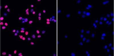
In Western blot this antibody detects ~17 kDa protein representing phosphorylated H2AX in gamma irradiated HeLa cell lysate. In immunofluorescence procedures, recognizes phosphorylated H2AX in gamma irradiated HeLa cells. ELISA of phosphorylated H2AX can also be performed. Used in IHC to successfully detect H2A.X pSer140 in postnatal mouse lung section.
Marker
DNA Double-strand break marker
Clonality
Monoclonal
Host
Mouse
Isotype
IgG1 kappa
Theoretical MW
15 kDa.
Disclaimer note: The observed molecular weight of the protein may vary from the listed predicted molecular weight due to post translational modifications, post translation cleavages, relative charges, and other experimental factors.
Disclaimer note: The observed molecular weight of the protein may vary from the listed predicted molecular weight due to post translational modifications, post translation cleavages, relative charges, and other experimental factors.
Scientific Data Images for Histone H2AX [p Ser139] Antibody (3F2)
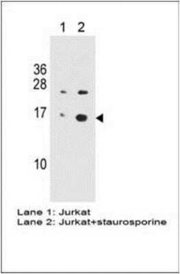
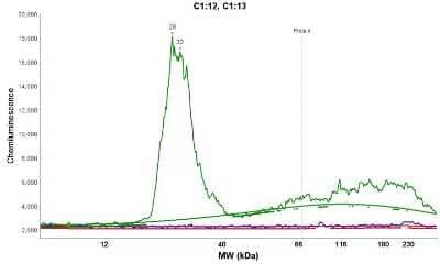
Simple Western: Histone H2AX [p Ser139] Antibody (3F2) [NB100-74435] - Simple Western lane view shows a specific band for Histone H2AX [p Ser139] in 0.2 mg/ml of Jurkat lysate(s). This experiment was performed under reducing conditions using the 12 - 230 kDa separation system.
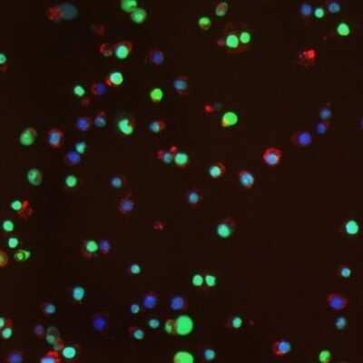
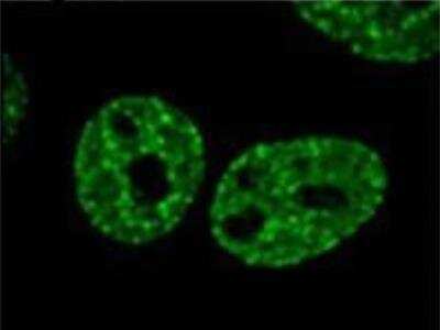
Immunocytochemistry/Immunofluorescence: Histone H2AX [p Ser139] Antibody (3F2) [NB100-74435] - Staining using NB100-74435, treatment with paraquat and iron induces MnSOD and Phosphorylation of H2AX in RAW 264.7 macrophages. Cells were treated for 20 hours with paraquat (500 uM) and iron (200 ug/ml) and stained with anti-Phospho-H2AX antibody.
Applications for Histone H2AX [p Ser139] Antibody (3F2)
Application
Recommended Usage
ELISA
1:100 - 1:2000
Flow Cytometry
1 ug/million cells
Immunocytochemistry/ Immunofluorescence
2 - 4 ug/ml
Immunohistochemistry
1:10 - 1:500
Immunohistochemistry-Paraffin
1:10 - 1:500
Simple Western
10 ug/ml
Western Blot
1 ug/ml
Application Notes
In WB: Detects an approx. 17 kDa protein representing phosphorylated H2AX in gamma irradiate Hela cell lysate.
In Simple Western only 10 - 15 uL of the recommended dilution is used per data point.
See Simple Western Antibody Database for Simple Western validation: Tested in Jurkat lysate, separated by Size, antibody dilution of 10 ug/mL, apparent MW was 28 kDa. Separated by Size-Wes, Sally Sue/Peggy Sue.
In Simple Western only 10 - 15 uL of the recommended dilution is used per data point.
See Simple Western Antibody Database for Simple Western validation: Tested in Jurkat lysate, separated by Size, antibody dilution of 10 ug/mL, apparent MW was 28 kDa. Separated by Size-Wes, Sally Sue/Peggy Sue.
Reviewed Applications
Read 2 reviews rated 5 using NB100-74435 in the following applications:
Formulation, Preparation, and Storage
Purification
Protein G purified
Formulation
PBS with 1 mg/ml BSA
Preservative
0.05% Sodium Azide
Concentration
1 mg/ml
Shipping
The product is shipped with polar packs. Upon receipt, store it immediately at the temperature recommended below.
Stability & Storage
Store at -20C. Avoid freeze-thaw cycles.
Background: Histone H2AX
References
1. Palla, V. V., Karaolanis, G., Katafigiotis, I., Anastasiou, I., Patapis, P., Dimitroulis, D., & Perrea, D. (2017). gamma-H2AX: Can it be established as a classical cancer prognostic factor?. Tumour biology : the journal of the International Society for Oncodevelopmental Biology and Medicine. https://doi.org/10.1177/1010428317695931
2. Kuo, L. J., & Yang, L. X. (2008). Gamma-H2AX - a novel biomarker for DNA double-strand breaks. In vivo (Athens, Greece).
3. Kinner, A., Wu, W., Staudt, C., & Iliakis, G. (2008). Gamma-H2AX in recognition and signaling of DNA double-strand breaks in the context of chromatin. Nucleic acids research. https://doi.org/10.1093/nar/gkn550
4. Redon, C. E., Weyemi, U., Parekh, P. R., Huang, D., Burrell, A. S., & Bonner, W. M. (2012). gamma-H2AX and other histone post-translational modifications in the clinic. Biochimica et biophysica acta. https://doi.org/10.1016/j.bbagrm.2012.02.021
5. H2AX: Uniprot (P16104)
Additional Histone H2AX Products
Product Documents for Histone H2AX [p Ser139] Antibody (3F2)
Product Specific Notices for Histone H2AX [p Ser139] Antibody (3F2)
Licensed to Novus Biologicals LLC under U.S. Patent Nos. 6,362,317 and 6,884,873.
This product is for research use only and is not approved for use in humans or in clinical diagnosis. Primary Antibodies are guaranteed for 1 year from date of receipt.
Loading...
Loading...
Loading...
Loading...
Loading...

![Immunocytochemistry/ Immunofluorescence: Histone H2AX [p Ser139] Antibody (3F2) [NB100-74435] - Histone H2AX [p Ser139] Antibody (3F2)](https://resources.bio-techne.com/images/products/nb100-74435_mouse-monoclonal-histone-h2ax-p-ser139-antibody-3f2-31020241534396.jpg)