Human Akt2 Antibody
R&D Systems, part of Bio-Techne | Catalog # MAB23152


Conjugate
Catalog #
Key Product Details
Species Reactivity
Validated:
Human
Cited:
Human
Applications
Validated:
CyTOF-ready, Immunohistochemistry, Intracellular Staining by Flow Cytometry
Cited:
Immunohistochemistry-Paraffin
Label
Unconjugated
Antibody Source
Monoclonal Mouse IgG1 Clone # 302501
Product Specifications
Immunogen
S. frugiperda insect ovarian cell line Sf 21-derived recombinant human Akt2
Asn2-Glu481
Accession # P31751
Asn2-Glu481
Accession # P31751
Specificity
Detects human Akt2. Using direct ELISA, this antibody does not detect recombinant human (rh) Akt1 or rhAkt3.
Clonality
Monoclonal
Host
Mouse
Isotype
IgG1
Scientific Data Images for Human Akt2 Antibody
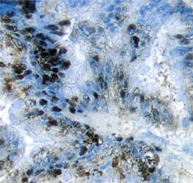
Akt2 in Human Pancreatic Cancer Tissue.
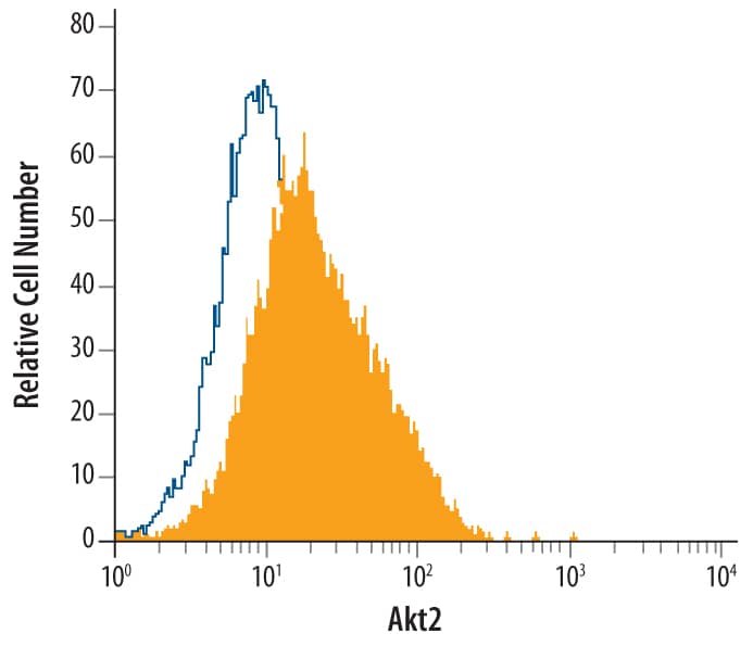
Akt2 was detected in immersion fixed paraffin-embedded sections of human pancreatic cancer tissue using 25 µg/mL Mouse Anti-Human Akt2 Monoclonal Antibody (Catalog # MAB23152) overnight at 4 °C. Tissue was stained with the Anti-Mouse HRP-DAB Cell & Tissue Staining Kit (brown; Catalog # CTS002) and counterstained with hematoxylin (blue). Specific labeling was localized to the cytoplasm in epithelial cells. View our protocol for Chromogenic IHC Staining of Paraffin-embedded Tissue Sections.Detection of Akt2 in MCF‑7 Human Cell Line by Flow Cytometry.
MCF-7 human breast cancer cell line was stained with Mouse Anti-Human Akt2 Monoclonal Antibody (Catalog # MAB23152, filled histogram) or isotype control antibody (Catalog # MAB002, open histogram), followed by Phycoerythrin-conjugated Anti-Mouse IgG F(ab')2Secondary Antibody (Catalog # F0102B). To facilitate intracellular staining, cells were fixed with paraformaldehyde and permeabilized with saponin.Applications for Human Akt2 Antibody
Application
Recommended Usage
CyTOF-ready
Ready to be labeled using established conjugation methods. No BSA or other carrier proteins that could interfere with conjugation.
Immunohistochemistry
8-25 µg/mL
Sample: Immersion fixed paraffin-embedded sections of human pancreatic cancer tissue
Sample: Immersion fixed paraffin-embedded sections of human pancreatic cancer tissue
Intracellular Staining by Flow Cytometry
2.5 µg/106 cells
Sample: MCF-7 human breast cancer cell line fixed with paraformaldehyde and permeabilized with saponin
Sample: MCF-7 human breast cancer cell line fixed with paraformaldehyde and permeabilized with saponin
Formulation, Preparation, and Storage
Purification
Protein A or G purified from hybridoma culture supernatant
Reconstitution
Reconstitute at 0.5 mg/mL in sterile PBS. For liquid material, refer to CoA for concentration.
Formulation
Lyophilized from a 0.2 μm filtered solution in PBS with Trehalose. *Small pack size (SP) is supplied either lyophilized or as a 0.2 µm filtered solution in PBS.
Shipping
Lyophilized product is shipped at ambient temperature. Liquid small pack size (-SP) is shipped with polar packs. Upon receipt, store immediately at the temperature recommended below.
Stability & Storage
Use a manual defrost freezer and avoid repeated freeze-thaw cycles.
- 12 months from date of receipt, -20 to -70 °C as supplied.
- 1 month, 2 to 8 °C under sterile conditions after reconstitution.
- 6 months, -20 to -70 °C under sterile conditions after reconstitution.
Background: Akt2
The serine/threonine kinase Akt, also known as protein kinase B (PKB), is a central regulator of such diverse cellular processes as glucose uptake, cell cycle progression, and apoptosis. In mammals, three highly homologous members define the Akt family: Akt1 (PKB alpha), Akt2 (PKB beta), and Akt3 (PKB gamma). Akt2 is expressed predominantly in insulin target tissues such as liver, skeletal muscle, and fat.
Long Name
v-Akt Murine Thymoma Viral Oncogene Homolog 2
Alternate Names
PKB beta, RAC-beta
Gene Symbol
AKT2
UniProt
Additional Akt2 Products
Product Documents for Human Akt2 Antibody
Product Specific Notices for Human Akt2 Antibody
For research use only
Loading...
Loading...
Loading...
Loading...
Loading...
