Human Apolipoprotein E/ApoE Antibody
R&D Systems, part of Bio-Techne | Catalog # AF4144

Key Product Details
Species Reactivity
Validated:
Cited:
Applications
Validated:
Cited:
Label
Antibody Source
Product Specifications
Immunogen
Lys19-His317
Accession # P02649
Specificity
Clonality
Host
Isotype
Scientific Data Images for Human Apolipoprotein E/ApoE Antibody
Detection of Human Apolipoprotein E/ApoE by Western Blot.
Western blot shows lysates of human plasma. PVDF membrane was probed with 1 µg/mL of Goat Anti-Human Apolipoprotein E/ApoE Antigen Affinity-purified Polyclonal Antibody (Catalog # AF4144) followed by HRP-conjugated Anti-Goat IgG Secondary Antibody (Catalog # HAF019). A specific band was detected for Apolipoprotein E/ApoE at approximately 38 kDa (as indicated). This experiment was conducted under reducing conditions and using Immunoblot Buffer Group 8.Detection of Human Apolipoprotein E/ ApoE by Western Blot.
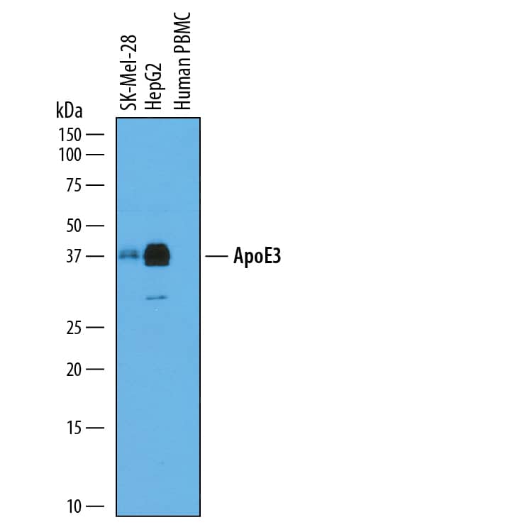
Western blot shows conditioned media from SK-Mel-28 human malignant melanoma cell line, HepG2 human hepatocellular carcinoma cell line, and human peripheral blood mononuclear cells (PBMC). PVDF membrane was probed with 0.5 µg/mL of Goat Anti-Human Apolipoprotein E/ApoE Antigen Affinity-purified Polyclonal Antibody (Catalog # AF4144) followed by HRP-conjugated Anti-Goat IgG Secondary Antibody (Catalog # HAF109). A specific band was detected for Apolipoprotein E/ApoE at approximately 38 kDa (as indicated). This experiment was conducted under reducing conditions and using Immunoblot Buffer Group 1.Detection of Human Apolipoprotein E/ ApoE by Simple WesternTM.
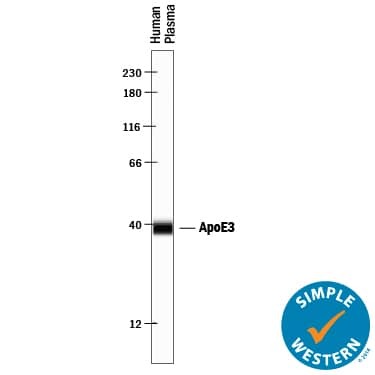
Simple Western lane view shows human plasma, loaded at 1:10 dilution. A specific band was detected for Apolipoprotein E/ApoE at approximately 39 kDa (as indicated) using 10 µg/mL of Goat Anti-Human Apolipoprotein E/ApoE Antigen Affinity-purified Polyclonal Antibody (Catalog # AF4144) followed by 1:50 dilution of HRP-conjugated Anti-Goat IgG Secondary Antibody (Catalog # HAF109). This experiment was conducted under reducing conditions and using the 12-230 kDa separation system.Applications for Human Apolipoprotein E/ApoE Antibody
Simple Western
Sample: Human plasma
Western Blot
Sample: Human plasma, SK‑Mel‑28 human malignant melanoma cell line, HepG2 human hepatocellular carcinoma cell line, and human peripheral blood mononuclear cells (PBMC)
Formulation, Preparation, and Storage
Purification
Reconstitution
Formulation
Shipping
Stability & Storage
- 12 months from date of receipt, -20 to -70 °C as supplied.
- 1 month, 2 to 8 °C under sterile conditions after reconstitution.
- 6 months, -20 to -70 °C under sterile conditions after reconstitution.
Background: Apolipoprotein E/ApoE
ApoE is a major protein component of serum LDL, VLDL, HDL, and chylomicrons. It is produced predominantly by hepatocytes, macrophages, and non-neuronal cells in the CNS. ApoE-containing particles transport triglycerides and cholesterol to peripheral tissues for cellular uptake and catabolism (1‑4). Mature human ApoE is a 37 kDa glycoprotein that consists of an N-terminal domain composed of four bundled alpha-helices, plus a hinge region and an extended alpha-helical C-terminal domain (2, 5). Its amphipathic nature and flexible structure enables it to adopt dramatically different conformations upon lipid association (2). ApoE is monomeric in lipid particles, although it forms oligomers when lipid-free (6). ApoE3 is the most abundant of the three common alleles in human; ApoE2 and ApoE4 differ by single aa substitutions (1). Mature human ApoE shares 71% aa sequence identity with mouse and rat ApoE. LDL receptor family proteins preferentially bind and internalize the lipid-bound form of ApoE with the exception of VLDLR which also efficiently internalizes lipid-free ApoE (7, 8). Lipoprotein uptake is facilitated by the initial binding of ApoE to cell surface heparan sulfate proteoglycans (HSPG) (9). Receptor/HSPG binding and lipid interactions primarily involve the N- and C-terminal regions of ApoE, respectively (2). Recycled lipid-free ApoE is formed into HDL particles through interactions with the lipid transporter ABCA1 (10). High cellular sterol content activates the nuclear hormone receptor LXR which promotes increased ApoE synthesis and increased sterol efflux, while low sterol content induces LDL R expression with increased sterol uptake and decreased ApoE production (11). ApoE3 dampens the TNF-alpha induced inflammatory response in vascular endothelial cells (12). In the CNS, ApoE blocks production of the amyloid A beta peptide by inhibiting the gamma-secretase cleavage of APP (13). It also complexes with A beta and promotes A beta internalization via LRP2 (14, 15).
References
- Martins, I.J. et al. (2006) Mol. Pschiatry 11:721.
- Hatters, D.M. et al. (2006) Trends Biochem. Sci. 31:445.
- Heeren, J. et al. (2006) Arterioscler. Thromb. Vasc. Biol. 26:442.
- Mahley, R.W. et al. (1984) J. Lipid. Res. 25:1277.
- Zannis, V.I. et al. (1984) J. Biol. Chem. 259:5495.
- Perugini, M.A. et al. (2000) J. Biol. Chem. 275:36758.
- Ruiz, J. et al. (2005) J. Lipid Res. 46:1721.
- Chroni, A. et al. (2005) Biochemistry 44:13132.
- Futamura, M. et al. (2005) J. Biol. Chem. 280:5414.
- Krimbou, L. et al. (2004) J. Lipid. Res. 45:839.
- Lucic, D. et al. (2007) J. Lipid Res. 48:366.
- Mullick, A.E. et al. (2007) Arterioscler. Thromb. Vasc. Biol. 27:339.
- Irizarry, M.C. et al. (2004) J. Neurochem. 90:1132.
- Naslund, J. et al. (1995) Neuron 15:219.
- Zerbinatti, C.V. et al. (2006) J. Biol. Chem. 281:36180.
Alternate Names
Gene Symbol
UniProt
Additional Apolipoprotein E/ApoE Products
Product Documents for Human Apolipoprotein E/ApoE Antibody
Product Specific Notices for Human Apolipoprotein E/ApoE Antibody
For research use only


