Human ARX Antibody
R&D Systems, part of Bio-Techne | Catalog # AF7068

Key Product Details
Species Reactivity
Validated:
Cited:
Applications
Validated:
Cited:
Label
Antibody Source
Product Specifications
Immunogen
Ser2-Ala100
Accession # Q96QS3
Specificity
Clonality
Host
Isotype
Scientific Data Images for Human ARX Antibody
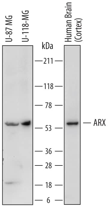
Detection of Human ARX by Western Blot.
Western blot shows lysates of U-87 MG human glioblastoma/astrocytoma cell line, U-118-MG human glioblastoma/astrocytoma cell line, and human brain (cortex) tissue. PVDF membrane was probed with 2 µg/mL of Sheep Anti-Human ARX Antigen Affinity-purified Polyclonal Antibody (Catalog # AF7068) followed by HRP-conjugated Anti-Sheep IgG Secondary Antibody (Catalog # HAF016). A specific band was detected for ARX at approximately 58 kDa (as indicated). This experiment was conducted under reducing conditions and using Immunoblot Buffer Group 1.ARX in U‑87 MG Human Cell Line.
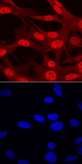
ARX was detected in immersion fixed U-87 MG human glioblastoma/astrocytoma cell line using Sheep Anti-Human ARX Antigen Affinity-purified Polyclonal Antibody (Catalog # AF7068) at 10 µg/mL for 3 hours at room temperature. Cells were stained using the NorthernLights™ 557-conjugated Anti-Sheep IgG Secondary Antibody (red, upper panel; Catalog # NL010) and counterstained with DAPI (blue, lower panel). Specific staining was localized to nuclei. View our protocol for Fluorescent ICC Staining of Cells on Coverslips.ARX in Human Pancreas.
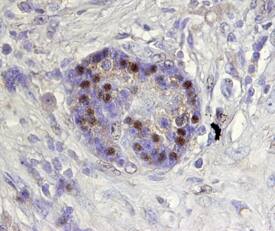
ARX was detected in immersion fixed paraffin-embedded sections of human pancreas using Sheep Anti-Human ARX Antigen Affinity-purified Polyclonal Antibody (Catalog # AF7068) at 15 µg/mL overnight at 4 °C. Tissue was stained using the Anti-Sheep HRP-DAB Cell & Tissue Staining Kit (brown; Catalog # CTS019) and counterstained with hematoxylin (blue). Specific staining was localized to nuclei of pancreatic islets. View our protocol for Chromogenic IHC Staining of Paraffin-embedded Tissue Sections.Applications for Human ARX Antibody
Immunocytochemistry
Sample: Immersion fixed U‑87 MG human glioblastoma/astrocytoma cell line
Immunohistochemistry
Sample: Immersion fixed paraffin-embedded sections of human pancreas
Western Blot
Sample: U‑87 MG human glioblastoma/astrocytoma cell line, U‑118‑MG human glioblastoma/astrocytoma cell line, and human brain (cortex) tissue
Formulation, Preparation, and Storage
Purification
Reconstitution
Formulation
Shipping
Stability & Storage
- 12 months from date of receipt, -20 to -70 °C as supplied.
- 1 month, 2 to 8 °C under sterile conditions after reconstitution.
- 6 months, -20 to -70 °C under sterile conditions after reconstitution.
Background: ARX
ARX (Aristaless-Related homeoboX protein) is a 56-62 kDa member of the aristaless subfamily, paired homeobox family of proteins. In the nervous system, it is expressed in embryonic telencephalon plus adult cortical GABAergic neurons and differentiating neuronal stem cells; in the pancreas, it promotes the differentiation of alpha-cells at the expense of beta-cells. Human ARX is a 562 amino acid (aa) transcriptional repressor. It contains an octapeptide motif that binds Groucho proteins (aa 27‑34), an NLS (aa 82-89), one Ala-rich region (aa 100-155), an acidic/Glu-rich region (aa 224-253), a DNA-binding homeodomain with an embedded NLS (aa 328‑387), one Pro-rich segment that binds CtBP1 (aa 395-459) and a C-terminal Aristaless domain (aa 530-543). There are mutations that show poly-Ala expansions in the N-terminus (aa 115-155), often associated with developmental abnormalities. Over aa 1-100, human ARX shares 97% aa identity with mouse ARX.
Long Name
Alternate Names
Gene Symbol
UniProt
Additional ARX Products
Product Documents for Human ARX Antibody
Product Specific Notices for Human ARX Antibody
For research use only


