Human BAFF/BLyS/TNFSF13B Antibody
R&D Systems, part of Bio-Techne | Catalog # AF124


Key Product Details
Validated by
Species Reactivity
Validated:
Cited:
Applications
Validated:
Cited:
Label
Antibody Source
Product Specifications
Immunogen
Ala134-Leu285
Accession # Q9Y275
Specificity
Clonality
Host
Isotype
Endotoxin Level
Scientific Data Images for Human BAFF/BLyS/TNFSF13B Antibody
Cell Proliferation Induced By BAFF/BLyS/TNFSF13B and Neutralization by Human BAFF/BLyS/TNFSF13B Antibody.
In the presence of Goat F(ab')2 Anti-mouse IgM (1 µg/mL), Recombinant Human BAFF/BLyS/TNFSF13B (Catalog # 2149-BF) stimulates proliferation in mouse B cells in a dose-dependent manner (orange line). Under these conditions, proliferation elicited by Recombinant Human BAFF/BLyS/TNFSF13B (5 ng/mL) is neutralized (green line) by increasing concentrations of Goat Anti-Human BAFF/BLyS/TNFSF13B Antigen Affinity-purified Polyclonal Antibody (Catalog # AF124). The ND50 is typically 3-12 ng/mL.BAFF/BLyS/TNFSF13B in Human Spleen.
BAFF/BLyS/TNFSF13B was detected in formalin fixed paraffin-embedded sections of human spleen using 15 µg/mL Goat Anti-Human BAFF/BLyS/TNFSF13B Antigen Affinity-purified Polyclonal Antibody (Catalog # AF124) overnight at 4 °C. Tissue was stained with the Anti-Goat HRP-DAB Cell & Tissue Staining Kit (brown; Catalog # CTS008) and counterstained with hematoxylin (blue). View our protocol for Chromogenic IHC Staining of Paraffin-embedded Tissue Sections.Detection of Human BAFF/BLyS/TNFSF13B by Immunocytochemistry/ Immunofluorescence
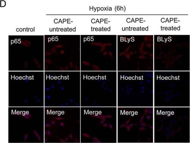
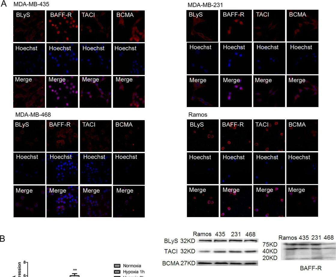
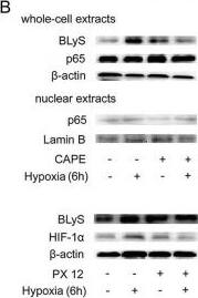
Role of p65 activation in BLyS up-regulation. (A) HIF-1 alpha and p65 protein levels in MDA-MB-435 in hypoxic conditions for different time points by Western Blotting. (B) CAPE(50 μM)and PX 12 (10 μM) were used to determine the roles of p65 and HIF-1 alpha in the regulation of BLyS expression by Western Blotting. The cells were treated with or without inhibitor in normoxic or hypoxic conditions for 6 h. (C) Effects of CAPE(50 μM)and PX 12 (10 μM) on BLyS promoter activity. Data were average luciferase activities of three independent transfections with ± SD. *, P < 0.05, vs pGL3-Basic/BP. (D) Localization of p65 protein and expression level of BLyS by immunofluorescence. MDA-MB-435 cells were challenged with CAPE (50 μM) for 6 h (original magnification 200 ×). Image collected and cropped by CiteAb from the following open publication (https://jeccr.biomedcentral.com/articles/10.1186/1756-9966-31-31), licensed under a CC-BY license. Not internally tested by R&D Systems.Applications for Human BAFF/BLyS/TNFSF13B Antibody
Immunohistochemistry
Sample: Formalin fixed paraffin-embedded sections of human spleen
Western Blot
Sample: Recombinant Human BAFF/BLyS/TNFSF13B (Catalog # 2149-BF)
Neutralization
Reviewed Applications
Read 2 reviews rated 5 using AF124 in the following applications:
Formulation, Preparation, and Storage
Purification
Reconstitution
Formulation
Shipping
Stability & Storage
- 12 months from date of receipt, -20 to -70 °C as supplied.
- 1 month, 2 to 8 °C under sterile conditions after reconstitution.
- 6 months, -20 to -70 °C under sterile conditions after reconstitution.
Background: BAFF/BLyS/TNFSF13B
BAFF (also known as TALL-1, BLyS, and THANK) is a type II transmembrane glycoprotein belonging to the TNF superfamily and has been designated as TNF superfamily member 13B (TNFSF13B). Human BAFF is a 285 amino acid (aa) protein consisting of a 218 aa extracellular domain, a 21 aa transmembrane region and a 46 aa cytoplasmic tail (1, 2). BAFF has the typical structural characteristics of the TNF superfamily ligands. It is a homotrimeric protein having the structurally conserved motif known as TNF homology domain (3, 4). A higher ordered structure composed of a cluster of trimeric units resembling the structure of a viral capsid has also been reported (4). Human BAFF may be shed from the cell surface by proteolytic cleavage between R133 and Ala134 to yield a soluble form of the protein that is detectable in serum (1, 5). Within the TNF superfamily BAFF shares the highest homology (48%) with APRIL (1). BAFF shares with APRIL the ability to bind to BCMA and TACI and also binds specifically to BAFF receptor (BAFF R, also known as BR3 or TNFSFR13C), which is the principal BAFF receptor (6‑8). All three receptors are type III transmembrane proteins that are expressd in B cells. BAFF and APRIL can form active heteromers that bind to TACI (9). BAFF is expressed in peripheral blood mononuclear cells, in spleen and lymph nodes. Its expression in resting monocytes is up-regulated by IFN-alpha, IFN-beta, LPS and IL-10. BAFF provides critical survival signals to a subset of B cells with intermediate maturation status (T2 B cells) during the immune response (10). BAFF also plays an important role in the development of lymphoid tissue and enhances the survival of activated memory B cells (7, 11). Human and mouse BAFF share 86% aa sequence identity (1).
References
- Schneider, P. et al. (1999) J. Exp. Med. 189:1747.
- Mukhopadhyay, A. et al. (1999) J. Biol. Chem. 274:15978.
- Karpusas, M. et al. (2002) J. Mol. Biol. 315:1145.
- Liu, Y. et al. (2002) Cell 108:383.
- Cheema, G.S. et al. (2001) Arthr. Rheum. 44:1313.
- Marsters, S.A. et al. (2000) Curr. Biol. 10:785.
- Thompson, J.S. et al. (2001) Science 293:2108.
- Ng, L.G. et al. (2004) J. Immunol. 173:807.
- Roschke, V. et al. (2002) J. Immunol. 169:4314.
- Batten, M. et al. (2000) J. Exp. Med. 192:1453.
- Avery, D.T. et al. (2003) J. Clin. Invest. 112:286.
Long Name
Alternate Names
Gene Symbol
UniProt
Additional BAFF/BLyS/TNFSF13B Products
Product Documents for Human BAFF/BLyS/TNFSF13B Antibody
Product Specific Notices for Human BAFF/BLyS/TNFSF13B Antibody
For research use only


