Human BAFF/BLyS/TNFSF13B PE-conjugated Antibody
R&D Systems, part of Bio-Techne | Catalog # IC1241P
Clone 137314 was used by HLDA to establish CD designation


Conjugate
Catalog #
Key Product Details
Validated by
Biological Validation
Species Reactivity
Validated:
Human
Cited:
Human
Applications
Validated:
Flow Cytometry
Cited:
Flow Cytometry, Immunocytochemistry
Label
Phycoerythrin (Excitation = 488 nm, Emission = 565-605 nm)
Antibody Source
Monoclonal Mouse IgG1 Clone # 137314
Product Specifications
Immunogen
E. coli-derived recombinant human BAFF/BLyS/TNFSF13B
Ala81-Leu285
Accession # Q9Y275
Ala81-Leu285
Accession # Q9Y275
Specificity
Detects human BAFF/BLyS/TNFSF13B in direct ELISAs and Western blots. In Western blots, approximately 5% cross‑reactivity with recombinant human (rh) TL1A/TNSF15 is observed and no cross-reactivity with rhAPRIL, rhTNF-alpha, rhFas Ligand, rhGITR Ligand, rhLIGHT, rhTRAIL, rhTRANCE, or rhTWEAK is observed.
Clonality
Monoclonal
Host
Mouse
Isotype
IgG1
Scientific Data Images for Human BAFF/BLyS/TNFSF13B PE-conjugated Antibody
Detection of BAFF/BLyS/TNFSF13B in Human PBMCs by Flow Cytometry.
Human peripheral blood mononuclear cells (PBMCs) treated with PMA and Calcium Ionomycin (filled histogram) or resting (open histogram), were stained with Mouse Anti-Human BAFF/BLyS/TNFSF13B PE-conjugated Monoclonal Antibody (Catalog # IC1241P). To facilitate intracellular staining, cells were fixed with Flow Cytometry Fixation Buffer (FC004) and permeabilized with Flow Cytometry Permeabilization/Wash Buffer I (FC005). View our protocol for Staining Intracellular Molecules.Detection of BAFF/BLyS/TNFSF13B in THP-1 cells by Flow Cytometry
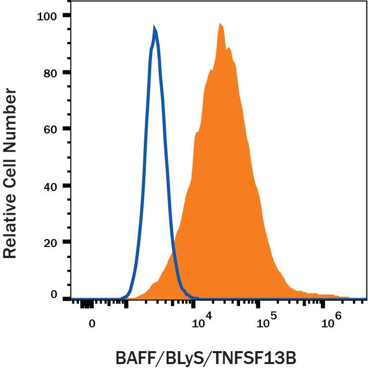
THP-1 cells were stained with Mouse Anti-Human BAFF/BLyS/TNFSF13B PE-conjugated Monoclonal Antibody (Catalog # IC1241P, filled histogram) or isotype control antibody (Catalog # IC002P, open histogram). View our protocol for Staining Membrane-associated Proteins.Applications for Human BAFF/BLyS/TNFSF13B PE-conjugated Antibody
Application
Recommended Usage
Flow Cytometry
0.25 µg/106 cells
Sample: THP-1 cells
Sample: THP-1 cells
Formulation, Preparation, and Storage
Purification
Protein A or G purified from hybridoma culture supernatant
Formulation
Supplied in a saline solution containing BSA and Sodium Azide.
Shipping
The product is shipped with polar packs. Upon receipt, store it immediately at the temperature recommended below.
Stability & Storage
Store the unopened product at 2 - 8° C. Do not use past expiration date. Protect from light.
Background: BAFF/BLyS/TNFSF13B
References
- Schneider, P. et al. (1999) J. Exp. Med. 189:1747.
- Mukhopadhyay, A. et al. (1999) J. Biol. Chem. 274:15978.
- Karpusas, M. et al. (2002) J. Mol. Biol. 315:1145.
- Liu, Y. et al. (2002) Cell 108:383.
- Cheema, G.S. et al. (2001) Arthr. Rheum. 44:1313.
- Marsters, S.A. et al. (2000) Curr. Biol. 10:785.
- Thompson, J.S. et al. (2001) Science 293:2108.
- Ng, L.G. et al. (2004) J. Immunol. 173:807.
- Roschke, V. et al. (2002) J. Immunol. 169:4314.
- Batten, M. et al. (2000) J. Exp. Med. 192:1453.
- Avery, D.T. et al. (2003) J. Clin. Invest. 112:286.
Long Name
B cell Activating Factor
Alternate Names
BLyS, CD257, TALL1, THANK, TNFSF13B, ZTNF4
Gene Symbol
TNFSF13B
UniProt
Additional BAFF/BLyS/TNFSF13B Products
Product Documents for Human BAFF/BLyS/TNFSF13B PE-conjugated Antibody
Product Specific Notices for Human BAFF/BLyS/TNFSF13B PE-conjugated Antibody
For research use only
Loading...
Loading...
Loading...
Loading...
Loading...
