Human Bcl-2 Minus C-Terminus Antibody
R&D Systems, part of Bio-Techne | Catalog # MAB827

Key Product Details
Validated by
Species Reactivity
Validated:
Cited:
Applications
Validated:
Cited:
Label
Antibody Source
Product Specifications
Immunogen
Met1-Asp211
Accession # P10415
Specificity
Clonality
Host
Isotype
Scientific Data Images for Human Bcl-2 Minus C-Terminus Antibody
Detection of Human Bcl‑2 Minus C-Terminus by Western Blot.
Western blot shows lysates of KG-1 human acute myelogenous leukemia cell line. PVDF membrane was probed with 0.1-0.5 µg/mL of Mouse Anti-Human Bcl-2 Minus C-Terminus Monoclonal Antibody (Catalog # MAB827) followed by HRP-conjugated Anti-Mouse IgG Secondary Antibody (Catalog # HAF007). A specific band was detected for Bcl-2 Minus C-Terminus at approximately 24 kDa (as indicated). This experiment was conducted under reducing conditions and using Immunoblot Buffer Group 1.Detection of Human Bcl‑2 by Simple WesternTM.
Simple Western lane view shows lysates of MCF‑7 human breast cancer cell line, loaded at 0.5 mg/mL. A specific band was detected for Bcl‑2 at approximately 24 kDa (as indicated) using 5 µg/mL of Mouse Anti-Human Bcl‑2 Minus C-Terminus Monoclonal Antibody (Catalog # MAB827). This experiment was conducted under reducing conditions and using the 12-230 kDa separation system.Detection of Human Bcl-2 by Simple Western
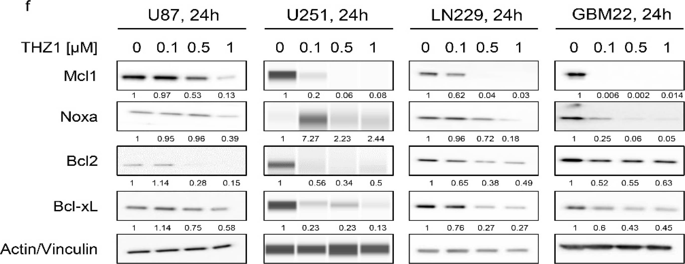
THZ1, suppresses Mcl-1 at the transcriptional level by disruption of its super-enhancer. (a) Real-time PCR analysis of Mcl1 mRNA levels in several GBM cells treated with vehicle or THZ1 for 24 h (n = 4). U87, U251, LN229, KNS42, and GBM22 cells were treated with 100 nM THZ1 while GBM14 was treated with 50 nM THZ1. Statistical significance was determined by two-tailed Student’s t-test; (b) U87 GBM cells were treated with DMSO or THZ1 100 nM for 24 h, subjected to CHIP with H3K27ac antibody and submitted for next generation sequencing. Super-enhancers were called using HOMER and depicted as a heatmap. The middle of each plot highlights the center of the super-enhancers (from −50 kb to 50 kb). The super enhancers are ranked by size and intensity levels are provided in the legend. The scale bar indicates the intensities. Blue depicts a high intensity level and red depicts a low intensity level; (c) A representation of global disruption of the super-enhancer landscape of U87 treated with DMSO or THZ1 100 nM in (b); (d) Shown are CHIP-seq (H3K27ac) tracks around the MCL1 locus (pile up values are indicated) in U87 treated with DMSO or THZ1 100 nM (super enhancer related to the Mcl-1 gene: chr1:150,601,879-150,630,909 (GRCh38/hg38)); (e) Standard western blots of cell lysates of U87 and GBM22 cells treated with increasing concentration of THZ1 for 24 h (pRpb1 corresponds to serine 5). Actin is used as a loading control. The protein expression levels were quantified using ImageJ (shown in cursive font); (f) Standard western blots or protein capillary electrophoresis of cell lysates of U87, U251, LN229, and GBM22 cells treated with increasing concentration of THZ1 for 24 h. Actin is used as a loading control in standard western blots and Vinculin is used as a loading control in protein capillary electrophoresis. The protein expression levels were quantified by using ImageJ (shown in cursive font). Uncropped blots are shown in Figure S10; (g) Shown are the protein expression levels of Mcl1, Noxa, Bcl2, and Bcl-xL following treatment with increasing concentration of THZ1 for 24 h in U87, U251, LN229, and GBM22 cells. FC: fold change. Shown are means and SD (n = 2–3). ***/**** p < 0.001. Image collected and cropped by CiteAb from the following publication (https://pubmed.ncbi.nlm.nih.gov/32752193), licensed under a CC-BY license. Not internally tested by R&D Systems.Applications for Human Bcl-2 Minus C-Terminus Antibody
Simple Western
Sample: MCF‑7 human breast cancer cell line
Western Blot
Sample: KG-1 human acute myelogenous leukemia cell line
Reviewed Applications
Read 3 reviews rated 3.7 using MAB827 in the following applications:
Formulation, Preparation, and Storage
Purification
Reconstitution
Formulation
Shipping
Stability & Storage
- 12 months from date of receipt, -20 to -70 °C as supplied.
- 1 month, 2 to 8 °C under sterile conditions after reconstitution.
- 6 months, -20 to -70 °C under sterile conditions after reconstitution.
Background: Bcl-2
Bcl-2 is a member of a family of proteins that regulates outer mitochondrial membrane permeability (1, 2). Bcl-2 is an anti-apoptotic member that prevents release of cytochrome c from the mitochondria intermembrane space into the cytosol. Bcl-2 is present on the outer mitochondrial membrane and is also found on other membranes in some cell types. Natural Bcl-2 contains a carboxyl-terminal mitochondria targeting sequence. Recombinant Bcl-2, missing the mitochondrial targeting sequence, maintains its ability to neutralize pro-apoptotic Bcl-2 family members. Neutralization by Bcl-2 appears to be through binding the BH3 region of pro-apoptotic Bcl-2 family members. This activity does not require the mitochondrial targeting sequence.
References
- Gross, A. et al. (1999) Genes and Develop. 13:1899.
- Kroemer, G. (1997) Nature Med. 3:614.
Long Name
Alternate Names
Gene Symbol
UniProt
Additional Bcl-2 Products
Product Documents for Human Bcl-2 Minus C-Terminus Antibody
Product Specific Notices for Human Bcl-2 Minus C-Terminus Antibody
For research use only


