Human beta-Galactosidase-1/GLB1 Antibody
R&D Systems, part of Bio-Techne | Catalog # AF6464

Key Product Details
Species Reactivity
Human
Applications
Simple Western, Western Blot
Label
Unconjugated
Antibody Source
Polyclonal Sheep IgG
Product Specifications
Immunogen
Chinese hamster ovary cell line CHO-derived recombinant human beta‑Galactosidase‑1/GLB1
Leu24-Val677
Accession # AAA51819
Leu24-Val677
Accession # AAA51819
Specificity
Detects human beta‑Galactosidase‑1/GLB1 in direct ELISAs and Western blots.
Clonality
Polyclonal
Host
Sheep
Isotype
IgG
Scientific Data Images for Human beta-Galactosidase-1/GLB1 Antibody
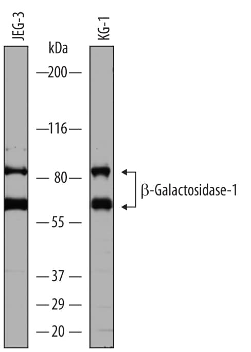
Detection of Human beta‑Galactosidase‑1/GLB1 by Western Blot.
Western blot shows lysates of JEG-3 human epithelial choriocarcinoma cell line and KG-1 human acute myelogenous leukemia cell line. PVDF membrane was probed with 1 µg/mL of Sheep Anti-Human beta-Galactosidase-1/GLB1 Antigen Affinity-purified Polyclonal Antibody (Catalog # AF6464) followed by HRP-conjugated Anti-Sheep IgG Secondary Antibody (Catalog # HAF016). Specific bands were detected for beta-Galactosidase-1/GLB1 at approximately 60 kDa and 80 kDa (as indicated). This experiment was conducted under reducing conditions and using Immunoblot Buffer Group 8.Detection of Human beta‑Galactosidase‑1/GLB1 by Simple WesternTM.
Simple Western lane view shows lysates of JEG-3 human epithelial choriocarcinoma cell line, loaded at 0.2 mg/mL. Specific bands were detected for beta-Galactosidase-1/GLB1 at approximately 71 & 110 kDa (as indicated) using 10 µg/mL of Sheep Anti-Human beta-Galactosidase-1/GLB1 Antigen Affinity-purified Polyclonal Antibody (Catalog # AF6464) followed by 1:50 dilution of HRP-conjugated Anti-Sheep IgG Secondary Antibody (Catalog # HAF016). This experiment was conducted under reducing conditions and using the 12-230 kDa separation system.Applications for Human beta-Galactosidase-1/GLB1 Antibody
Application
Recommended Usage
Simple Western
10 µg/mL
Sample: JEG‑3 human epithelial choriocarcinoma cell line
Sample: JEG‑3 human epithelial choriocarcinoma cell line
Western Blot
1 µg/mL
Sample: JEG‑3 human epithelial choriocarcinoma cell line and KG‑1 human acute myelogenous leukemia cell line
Sample: JEG‑3 human epithelial choriocarcinoma cell line and KG‑1 human acute myelogenous leukemia cell line
Formulation, Preparation, and Storage
Purification
Antigen Affinity-purified
Reconstitution
Sterile PBS to a final concentration of 0.2 mg/mL. For liquid material, refer to CoA for concentration.
Formulation
Lyophilized from a 0.2 μm filtered solution in PBS with Trehalose. *Small pack size (SP) is supplied either lyophilized or as a 0.2 µm filtered solution in PBS.
Shipping
Lyophilized product is shipped at ambient temperature. Liquid small pack size (-SP) is shipped with polar packs. Upon receipt, store immediately at the temperature recommended below.
Stability & Storage
Use a manual defrost freezer and avoid repeated freeze-thaw cycles.
- 12 months from date of receipt, -20 to -70 °C as supplied.
- 1 month, 2 to 8 °C under sterile conditions after reconstitution.
- 6 months, -20 to -70 °C under sterile conditions after reconstitution.
Background: beta-Galactosidase-1/GLB1
References
- Hofer, D. et al. (2009) Hum. Mutat. 30:1214.
- Brunetti-Pierri N, and Scaglia F. (2008) Mol. Genet. Metab. 94:391.
- Santamaria, R. et al. (2007) J. Lipid Res. 48:2275.
- Prat, C. (2008) Joint Bone Spine, 75:495.
- Zhang, S. et al. (2000) Biochem. J. 348:621.
- Pshezhetsky, A.V. and Ashmarina, M. (2001) Prog. Nucleic Acid Res. Mol. Biol. 69:81.
- Salvatore P, et al. (1998) J. Biol. Chem. 273:6319.
Alternate Names
betaGalactosidase1, EBP, ELNR1, GLB1, Lactase
Entrez Gene IDs
2720 (Human)
Gene Symbol
GLB1
UniProt
Additional beta-Galactosidase-1/GLB1 Products
Product Documents for Human beta-Galactosidase-1/GLB1 Antibody
Product Specific Notices for Human beta-Galactosidase-1/GLB1 Antibody
For research use only
Loading...
Loading...
Loading...
Loading...

