Human BMI-1 Antibody
R&D Systems, part of Bio-Techne | Catalog # MAB33341


Key Product Details
Species Reactivity
Validated:
Cited:
Applications
Validated:
Cited:
Label
Antibody Source
Product Specifications
Immunogen
Asp96-Gly326
Accession # P35226
Specificity
Clonality
Host
Isotype
Scientific Data Images for Human BMI-1 Antibody
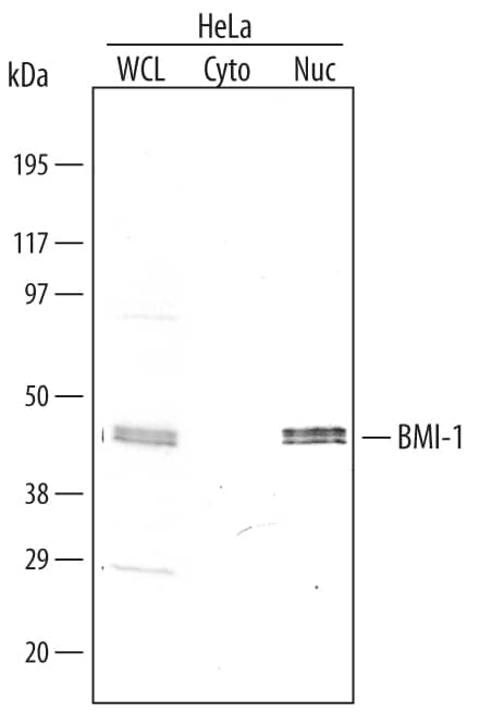
Detection of Human BMI-1 by Western Blot.
Western blot shows lysates of HeLa human cervical epithelial carcinoma cell line. Gels were loaded with 30 µg of whole cell lysate (WCL), 20 µg of cytoplasmic (Cyto), and 10 µg of nuclear extracts (Nuc). PVDF membrane was probed with 0.1 µg/mL Mouse Anti-Human BMI-1 Monoclonal Antibody (Catalog # MAB33341) followed by HRP-conjugated Anti-Mouse IgG Secondary Antibody (Catalog # HAF007). A specific band for BMI-1 was detected at approximately 45 kDa (as indicated). This experiment was conducted under reducing conditions and using Immunoblot Buffer Group 4.Detection of BMI‑1-regulated Genes by Chromatin Immunoprecipitation.
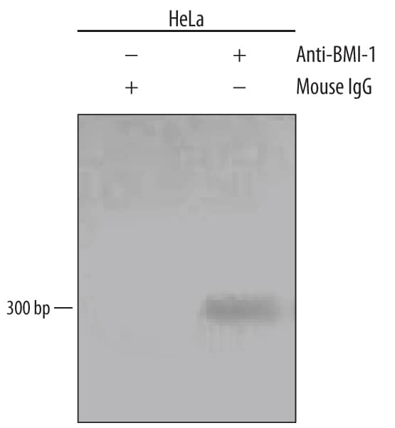
HeLa human cervical epithelial carcinoma cell line was fixed using formaldehyde, resuspended in lysis buffer, and sonicated to shear chromatin. BMI-1/DNA complexes were immunoprecipitated using 5 µg Mouse Anti-Human BMI-1 Monoclonal Antibody (Catalog # MAB33341) or control antibody (Catalog # MAB003) for 15 minutes in an ultrasonic bath, followed by Biotinylated Anti-Mouse IgG Secondary Antibody (Catalog # BAF007). Immunocomplexes were captured using 50 µL of MagCellect Streptavidin Ferrofluid (Catalog # MAG999) and DNA was purified using chelating resin solution. Thehoxc13promoter was detected by standard PCR.Detection of BMI-1 in HeLa Human Cell Line by Flow Cytometry.
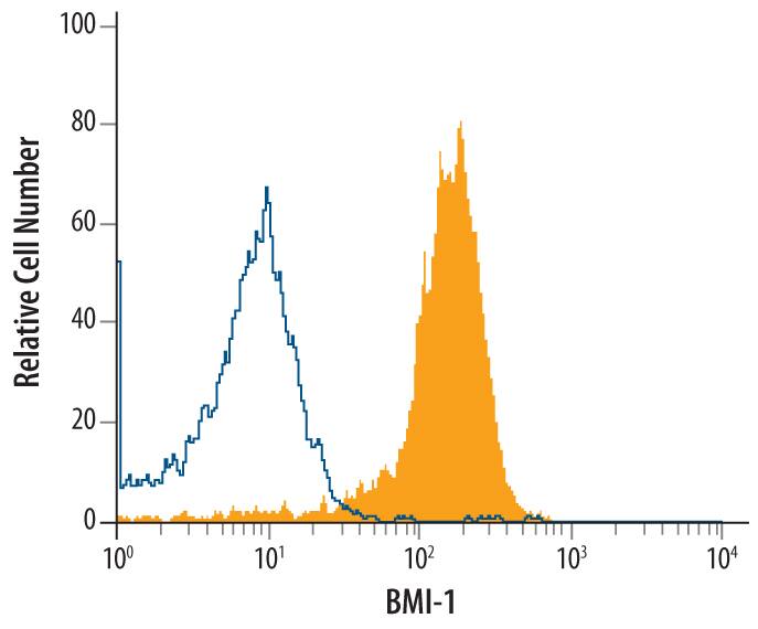
HeLa human cervical epithelial carcinoma cell line was stained with Mouse Anti-Human BMI-1 Monoclonal Antibody (Catalog # MAB33341, filled histogram) or isotype control antibody (Catalog # MAB003, open histogram), followed by Phycoerythrin-conjugated Anti-Mouse IgG F(ab')2Secondary Antibody (Catalog # F0102B). To facilitate intracellular staining, cells were fixed with paraformaldehyde and permeabilized with saponin.Applications for Human BMI-1 Antibody
Chromatin Immunoprecipitation (ChIP)
Sample: HeLa human cervical epithelial carcinoma cell line chromatin, hoxc13 promoter detected by standard PCR
CyTOF-ready
Intracellular Staining by Flow Cytometry
Sample: HeLa human cervical epithelial carcinoma cell line fixed with paraformaldehyde and permeabilized with saponin
Western Blot
Sample: HeLa human cervical epithelial carcinoma cell line
Reviewed Applications
Read 1 review rated 5 using MAB33341 in the following applications:
Formulation, Preparation, and Storage
Purification
Reconstitution
Formulation
Shipping
Stability & Storage
- 12 months from date of receipt, -20 to -70 °C as supplied.
- 1 month, 2 to 8 °C under sterile conditions after reconstitution.
- 6 months, -20 to -70 °C under sterile conditions after reconstitution.
Background: BMI-1
BMI-1 (B cell-specific Moloney-MLV integration site #1) is a 45 kDa protooncogene that is a class II member of the Polycomb group of genes. It participates in the formation of a large multimeric complex termed PRC1 that inhibits target gene transcription. Loss of BMI-1 function precludes stem cells from self-replicating. Human BMI-1 contains an N-terminal RING-finger domain (aa 17-56), an NLS (aa 81-95) and a C-terminal Pro/Ser-rich region (aa 251-326). Human BMI-1 shares 99%, 97%, 99% and 99% aa sequence identity with bovine, mouse, feline and canine BMI-1, respectively.
Long Name
Alternate Names
Gene Symbol
UniProt
Additional BMI-1 Products
Product Documents for Human BMI-1 Antibody
Product Specific Notices for Human BMI-1 Antibody
For research use only

