Human Cadherin-11 PE-conjugated Antibody
R&D Systems, part of Bio-Techne | Catalog # FAB17901P


Conjugate
Catalog #
Key Product Details
Species Reactivity
Human
Applications
Flow Cytometry
Label
Phycoerythrin (Excitation = 488 nm, Emission = 565-605 nm)
Antibody Source
Monoclonal Mouse IgG2A Clone # 667039
Product Specifications
Immunogen
Mouse myeloma cell line NS0-derived recombinant human Cadherin-11
Phe23-Thr617
Accession # AAA35622
Phe23-Thr617
Accession # AAA35622
Specificity
Detects human Cadherin-11 in direct ELISAs. In direct ELISAs, no cross-reactivity with recombinant human (rh) Cadherin-4, -6, -8, -12, -13,-17, rhE-Cadherin, rhN-Cadherin, rhP-Cadherin, or rhVE-Cadherin is observed.
Clonality
Monoclonal
Host
Mouse
Isotype
IgG2A
Scientific Data Images for Human Cadherin-11 PE-conjugated Antibody
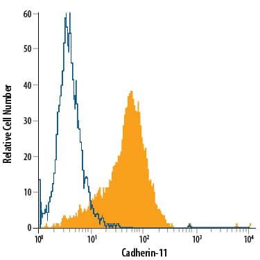
Detection of Cadherin‑11 in PC‑3 Human Cell Line by Flow Cytometry.
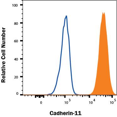
PC-3 human prostate cancer cell line was stained with Mouse Anti-Human Cadherin-11 PE-conjugated Monoclonal Antibody (Catalog # FAB17901P, filled histogram) or isotype control antibody (Catalog # IC003P, open histogram). Cells were stained in a buffer containing Ca2+and Mg2+. View our protocol for Staining Membrane-associated Proteins.Detection of Cadherin‑11 in NCI-H460 Human Cell Line by Flow Cytometry.
NCI-H460 human small cell lung cancer cell line was stained with Mouse Anti-Human Cadherin-11 PE-conjugated Monoclonal Antibody (Catalog # FAB17901P, filled histogram) or isotype control antibody (IC003P, open histogram). Cells were stained in a buffer containing Ca2+and Mg2+. View our protocol for Staining Membrane-associated Proteins.Applications for Human Cadherin-11 PE-conjugated Antibody
Application
Recommended Usage
Flow Cytometry
10 µL/106 cells
Sample: PC-3 human prostate cancer cell line and NCI-H460 human small cell lung cancer cell line stained in buffer containing Ca2+ and Mg2+
Sample: PC-3 human prostate cancer cell line and NCI-H460 human small cell lung cancer cell line stained in buffer containing Ca2+ and Mg2+
Formulation, Preparation, and Storage
Purification
Protein A or G purified from hybridoma culture supernatant
Formulation
Supplied in a saline solution containing BSA and Sodium Azide.
Shipping
The product is shipped with polar packs. Upon receipt, store it immediately at the temperature recommended below.
Stability & Storage
Protect from light. Do not freeze.
- 12 months from date of receipt, 2 to 8 °C as supplied.
Background: Cadherin-11
References
- Pokutta, S. and W.I. Weis (2007) Annu. Rev. Cell Dev. Biol. 23:237.
- Kimura, Y. et al. (1995) Dev. Biol. 169:347.
- McCusker, C. et al. (2009) Mol. Biol. Cell 20:78.
- Clendenon, S.G. et al. (2009) Dev. Dyn. 238:1909.
- Kii, I. et al. (2004) J. Bone Mineral Res. 19:1840.
- Tamura, D. et al. (2008) Int. J. Oncol. 33:17.
- Chu, K. et al. (2008) Mol. Cancer Res. 6:1259.
- Huang, C.-F. et al. (2010) Cancer Res. 70:4580.
- Valencia, X. et al. (2004) J. Exp. Med. 200:1673.
- Kiener, H.P. et al. (2009) Arthritis Rheum. 60:1305.
- Monahan, T.S. et al. (2007) J. Vasc. Surg. 45:581.
- Boscher, C. and R.-M. Mege (2008) Cell. Signal. 20:1061.
- Tanihara, H. et al. (1994) Cell Adhes. Commun. 2:15.
- Okazaki, M. et al. (1994) J. Biol. Chem. 269:12092.
- Kawaguchi, J. et al. (1999) J. Bone Mineral Res. 14:764.
Alternate Names
Cadherin11, CDH11, CDHOB, OB-Cadherin
Gene Symbol
CDH11
UniProt
Additional Cadherin-11 Products
Product Documents for Human Cadherin-11 PE-conjugated Antibody
Product Specific Notices for Human Cadherin-11 PE-conjugated Antibody
For research use only
Loading...
Loading...
Loading...
Loading...
Loading...
