Human Cadherin-13 Antibody
R&D Systems, part of Bio-Techne | Catalog # AF3264

Key Product Details
Species Reactivity
Validated:
Human
Cited:
Human, Mouse, Rat
Applications
Validated:
CyTOF-ready, Flow Cytometry, Immunocytochemistry, Immunohistochemistry, Simple Western, Western Blot
Cited:
Immunocytochemistry, Immunohistochemistry, Immunohistochemistry-Frozen, Immunoprecipitation, Western Blot
Label
Unconjugated
Antibody Source
Polyclonal Goat IgG
Product Specifications
Immunogen
Mouse myeloma cell line NS0-derived recombinant human Cadherin-13
Glu23-Ala692
Accession # P55290
Glu23-Ala692
Accession # P55290
Specificity
Detects human Cadherin-13 in direct ELISAs and Western blots. In direct ELISAs, approximately 100% cross-reactivity with recombinant mouse Cadherin-13 is observed, and less than 10% cross-reactivity with recombinant human (rh) N-Cadherin is observed, and less than 2% cross-reactivity with rhCadherin-8, rhCadherin-11, rhCadherin-17, rhE-Cadherin, rhP-Cadherin, rhVE-Cadherin and rhR-Cadherin is observed.
Clonality
Polyclonal
Host
Goat
Isotype
IgG
Scientific Data Images for Human Cadherin-13 Antibody
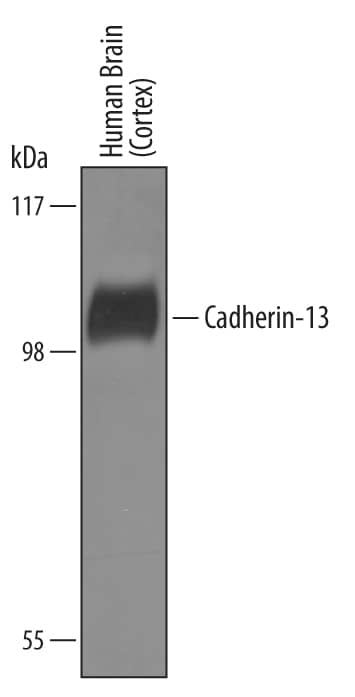
Detection of Human Cadherin‑13 by Western Blot.
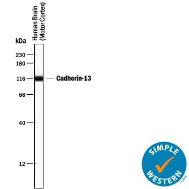
Western blot shows lysates of human brain (cortex) tissue. PVDF Membrane was probed with 1 µg/mL of Goat Anti-Human Cadherin-13 Antigen Affinity-purified Polyclonal Antibody (Catalog # AF3264) followed by HRP-conjugated Anti-Goat IgG Secondary Antibody (Catalog # HAF019). A specific band was detected for Cadherin-13 at approximately 105 kDa (as indicated). This experiment was conducted under reducing conditions and using Immunoblot Buffer Group 8.Cadherin‑13 in NCI-H460 Human Cell Line.
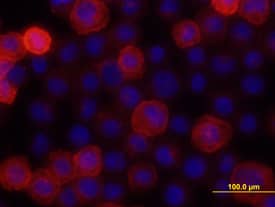
Cadherin-13 was detected in immersion fixed NCI-H460 human large cell lung carcinoma cell line using 10 µg/mL Goat Anti-Human Cadherin-13 Antigen Affinity-purified Polyclonal Antibody (Catalog # AF3264) for 3 hours at room temperature. Cells were stained with the NorthernLights™ 557-conjugated Anti-Goat IgG Secondary Antibody (red; Catalog # NL001) and counter-stained with DAPI (blue). View our protocol for Fluorescent ICC Staining of Cells on Coverslips.Cadherin‑13 in Human Skin.
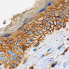
Cadherin-13 was detected in immersion fixed paraffin-embedded sections of human skin using Goat Anti-Human Cadherin-13 Antigen Affinity-purified Polyclonal Antibody (Catalog # AF3264) at 0.1 µg/mL overnight at 4 °C. Tissue was stained using the Anti-Goat HRP-DAB Cell & Tissue Staining Kit (brown; Catalog # CTS008) and counterstained with hematoxylin (blue). Specific staining was localized to plasma membranes of keratinocytes. View our protocol for Chromogenic IHC Staining of Paraffin-embedded Tissue Sections.Applications for Human Cadherin-13 Antibody
Application
Recommended Usage
CyTOF-ready
Ready to be labeled using established conjugation methods. No BSA or other carrier proteins that could interfere with conjugation.
Flow Cytometry
2.5 µg/106 cells
Sample: NCI-H460 human large cell lung carcinoma cell line
Sample: NCI-H460 human large cell lung carcinoma cell line
Immunocytochemistry
5-15 µg/mL
Sample: Immersion fixed NCI-H460 human large cell lung carcinoma cell line
Sample: Immersion fixed NCI-H460 human large cell lung carcinoma cell line
Immunohistochemistry
5-15 µg/mL
Sample: Immersion fixed paraffin-embedded sections of human skin
Sample: Immersion fixed paraffin-embedded sections of human skin
Simple Western
10 µg/mL
Sample: Human brain (motor cortex) tissue
Sample: Human brain (motor cortex) tissue
Western Blot
1 µg/mL
Sample: Human brain (cortex) tissue
Sample: Human brain (cortex) tissue
Formulation, Preparation, and Storage
Purification
Antigen Affinity-purified
Reconstitution
Reconstitute at 0.2 mg/mL in sterile PBS. For liquid material, refer to CoA for concentration.
Formulation
Lyophilized from a 0.2 μm filtered solution in PBS with Trehalose. *Small pack size (SP) is supplied either lyophilized or as a 0.2 µm filtered solution in PBS.
Shipping
Lyophilized product is shipped at ambient temperature. Liquid small pack size (-SP) is shipped with polar packs. Upon receipt, store immediately at the temperature recommended below.
Stability & Storage
Use a manual defrost freezer and avoid repeated freeze-thaw cycles.
- 12 months from date of receipt, -20 to -70 °C as supplied.
- 1 month, 2 to 8 °C under sterile conditions after reconstitution.
- 6 months, -20 to -70 °C under sterile conditions after reconstitution.
Background: Cadherin-13
References
- Tanihara, H. et al. (1994) Cell Adhes. Commun. 2:15.
- Philippova, M. et al. (2009) Cell. Signal. 21:1035.
Alternate Names
Cadherin13, CDH13, H-Cadherin, T-Cadherin
Gene Symbol
CDH13
UniProt
Additional Cadherin-13 Products
Product Documents for Human Cadherin-13 Antibody
Product Specific Notices for Human Cadherin-13 Antibody
For research use only
Loading...
Loading...
Loading...
Loading...
Loading...
