Human Cathepsin D Antibody
R&D Systems, part of Bio-Techne | Catalog # AF1014


Key Product Details
Species Reactivity
Validated:
Cited:
Applications
Validated:
Cited:
Label
Antibody Source
Product Specifications
Immunogen
Leu21-Leu412
Accession # P07339
Specificity
Clonality
Host
Isotype
Scientific Data Images for Human Cathepsin D Antibody
Detection of Human Cathepsin D by Western Blot.
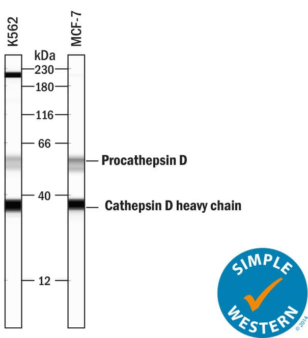
Western blot shows lysates of K562 human chronic myelogenous leukemia cell line. PVDF membrane was probed with 1 µg/mL of Goat Anti-Human Cathepsin D Antigen Affinity-purified Polyclonal Antibody (Catalog # AF1014) followed by HRP-conjugated Anti-Goat IgG Secondary Antibody (Catalog # HAF019). Specific bands were detected for Procathepsin D at approximately 45 kDa and Cathepsin D heavy chain 28 kDa (as indicated). This experiment was conducted under reducing conditions and using Immunoblot Buffer Group 1.Detection of Human Cathepsin D by Western Blot.
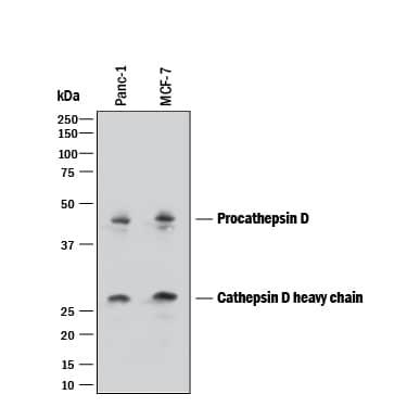
Western blot shows lysates of PANC-1 human pancreatic carcinoma cell line and MCF-7 human breast cancer cell line. PVDF membrane was probed with 1 µg/mL of Goat Anti-Human Cathepsin D Antigen Affinity-purified Polyclonal Antibody (Catalog # AF1014) followed by HRP-conjugated Anti-Goat IgG Secondary Antibody (Catalog # HAF017). Specific bands were detected for Procathepsin D at approximately 45 kDa and Cathepsin D heavy chain 28 kDa (as indicated). This experiment was conducted under reducing conditions and using Immunoblot Buffer Group 1.Cathepsin D in Human Lung Cancer Tissue.
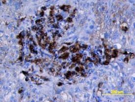
Cathepsin D was detected in immersion fixed paraffin-embedded sections of human lung cancer tissue using Goat Anti-Human Cathepsin D Antigen Affinity-purified Polyclonal Antibody (Catalog # AF1014) at 15 µg/mL overnight at 4 °C. Tissue was stained using the Anti-Goat HRP-DAB Cell & Tissue Staining Kit (brown; Catalog # CTS008) and counterstained with hematoxylin (blue). View our protocol for Chromogenic IHC Staining of Paraffin-embedded Tissue Sections.Applications for Human Cathepsin D Antibody
Immunohistochemistry
Sample: Immersion fixed paraffin-embedded sections of human breast and lung cancer tissue
Immunoprecipitation
Sample: Conditioned cell culture medium spiked with Recombinant Human Cathepsin D (Catalog # 1014-AS), see our available Western blot detection antibodies
Simple Western
Sample: K562 human chronic myelogenous leukemia cell line and MCF‑7 human breast cancer cell line
Western Blot
Sample: K562 human chronic myelogenous leukemia cell line, PANC‑1 human pancreatic carcinoma cell line, and MCF‑7 human breast cancer cell line
Formulation, Preparation, and Storage
Purification
Reconstitution
Formulation
Shipping
Stability & Storage
- 12 months from date of receipt, -20 to -70 °C as supplied.
- 1 month, 2 to 8 °C under sterile conditions after reconstitution.
- 6 months, -20 to -70 °C under sterile conditions after reconstitution.
Background: Cathepsin D
Cathepsin D is a lysosomal aspartic protease of the pepsin family (1). Human cathepsin D is synthesized as a precursor protein, consisting of a signal peptide (residues 1‑18), a propeptide (residues 19-64), and a mature chain (residues 65‑412) (2‑4). The mature chain can be processed further to the light (residues 65‑161) and heavy (residues 169‑412) chains. It is expressed in most cells and overexpressed in breast cancer cells (5). It is a major enzyme in protein degradation in lysosomes, and also involved in the presentation of antigenic peptides. Mice deficient in this enzyme showed a progressive atrophy of the intestinal mucosa, a massive destruction of lymphoid organs, and a profound neuronal ceroid lipofucinosis, indicating that cathepsin D is essential for proteolysis of proteins regulating cell growth and tissue homeostasis (6). Cathepsin D secreted from human prostate carcinoma cells are responsible for the generation of angiostatin, a potent endogeneous inhibitor of angiogenesis (6).
References
- Conner et al. in Handbook of Proteolytic Enzymes Barrett (1998) Academic Press, San Diego, p. 828.
- Faust, et al. (1985) Proc. Natl. Acad. Sci. USA 82:4910.
- Westley and May (1987) Nucl. Acid Res. 15:3773.
- Redecker, et al. (1991) DNA Cell Biol. 10:423.
- Rochefort, et al. (2000) Clin. Chim. Acta. 291:157.
- Tsukuba, et al. (2000) Mol. Cells 10:601.
Alternate Names
Gene Symbol
UniProt
Additional Cathepsin D Products
Product Documents for Human Cathepsin D Antibody
Product Specific Notices for Human Cathepsin D Antibody
For research use only


