Human CCL3/MIP-1 alpha Biotinylated Antibody Best Seller
R&D Systems, part of Bio-Techne | Catalog # BAF270


Key Product Details
Species Reactivity
Validated:
Cited:
Applications
Validated:
Cited:
Label
Antibody Source
Product Specifications
Immunogen
Ala27-Ala92
Accession # P10147
Specificity
Clonality
Host
Isotype
Scientific Data Images for Human CCL3/MIP-1 alpha Biotinylated Antibody
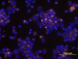
CCL3/MIP‑1 alpha in Human PBMCs.
CCL3/MIP-1a was detected in immersion fixed human peripheral blood mononuclear cells (PBMCs) stimulated with PHA and monensin using Human CCL3/MIP-1a Biotinylated Antigen Affinity-purified Polyclonal Antibody (Catalog # BAF270) at 10 µg/mL for 3 hours at room temperature. Cells were stained using the Northern-Lights™ 557-conjugated Streptavidin (yellow; Catalog # NL999) and counterstained with DAPI (blue). View our protocol for Fluorescent ICC Staining of Non-adherent Cells.Applications for Human CCL3/MIP-1 alpha Biotinylated Antibody
Immunocytochemistry
Sample: Immersion fixed human peripheral blood mononuclear cells (PBMCs) stimulated by PHA and monensin
Western Blot
Sample: Recombinant Human CCL3/MIP-1 alpha isoform LD78a (Catalog # 270-LD)
Human CCL3/MIP-1 alpha Sandwich Immunoassay
Formulation, Preparation, and Storage
Purification
Reconstitution
Formulation
Shipping
Stability & Storage
- 12 months from date of receipt, -20 to -70 °C as supplied.
- 1 month, 2 to 8 °C under sterile conditions after reconstitution.
- 6 months, -20 to -70 °C under sterile conditions after reconstitution.
Background: CCL3/MIP-1 alpha
The macrophage inflammatory proteins -1 alpha and -1 beta were originally co-purified from medium conditioned by an LPS-stimulated murine macrophage cell line. Human MIP-1 alpha refers to the products of several independently cloned cDNAs, including LD78, pL78, pAT464, and GOS19. These cDNAs all code for the same human protein that is a homologue of the murine MIP-1 alpha. Mature MIP-1 alpha and MIP-1 beta in both human and mouse share approximately 70% homology at the amino acid level. The MIP‑1 proteins are members of the beta (C-C) subfamily of chemokines.
Both MIP-1 alpha and MIP-1 beta are monocyte chemoattractants in vitro. Additionally, the MIP-1 proteins have been reported to have chemoattractant and adhesive effects on lymphocytes, with MIP-1 alpha and MIP-1 beta preferentially attracting CD8+ and CD4+ T cells, respectively. MIP-1 alpha has also been shown to attract B cells as well as eosinophils. MIP-1 proteins have been reported to have multiple effects on hematopoietic precursor cells and MIP-1 alpha has been identified as a stem cell inhibitory factor that can inhibit the proliferation of hematopoietic stem cells in vitro as well as in vivo. The functional receptor for MIP-1 alpha has been identified as CCR1 and CCR5.
References
- Menten, P. et al. (2002) Cytokine Growth Factor Rev. 13:455.
Alternate Names
Entrez Gene IDs
Gene Symbol
UniProt
Additional CCL3/MIP-1 alpha Products
Product Documents for Human CCL3/MIP-1 alpha Biotinylated Antibody
Product Specific Notices for Human CCL3/MIP-1 alpha Biotinylated Antibody
For research use only