Human CD163 PE-conjugated Antibody
R&D Systems, part of Bio-Techne | Catalog # FAB1607P


Key Product Details
Species Reactivity
Validated:
Cited:
Applications
Validated:
Cited:
Label
Antibody Source
Product Specifications
Immunogen
Gly46-Ser1050
Accession # Q86VB7
Specificity
Clonality
Host
Isotype
Scientific Data Images for Human CD163 PE-conjugated Antibody
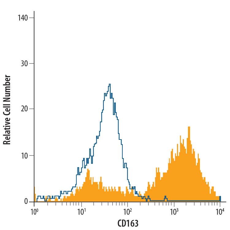
Detection of CD163 in Human Blood Monocytes by Flow Cytometry.
Human peripheral blood monocytes were stained with Mouse Anti-Human CD163 PE-conjugated Monoclonal Antibody (Catalog # FAB1607P, filled histogram) or isotype control antibody (Catalog # IC002P, open histogram). View our protocol for Staining Membrane-associated Proteins.Detection of Human CD163 by Flow Cytometry
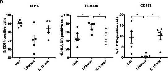
Phenotypic characterization of LPS- and IL-10-stimulated macrophages derived from human CD14+ peripheral blood monocytes. a Representative images of actin and tubulin stainings of LPS- and IL-10-stimulated macrophages polarized in absence of other external stimuli (mac) or in the presence of 10ng/ml LPS (LPSmac) or IL-10 (IL-10mac), respectively. F-actin was stained with Phalloidin-FITC (green), alpha–tubulin with a specific monoclonal antibody followed by incubation with AlexaFluor594 secondary antibody (red) and nuclei were counterstained with DAPI (blue). Scale bars represent 50 μm. b Morphological differences between macrophage populations were quantified by calculating the cell aspect ratio (quotient between cell major and minor axes) of actin/tubulin stained cells. Chart reflects measurements of at least 100 cells per donor from, at least, 3 distinct donors. Bars represent mean values and flags indicate standard deviations. c Cytokine production profile of LPS- and IL-10-stimulated macrophages. Cytokine concentration was measured by ELISA in conditioned media from distinct macrophage populations. Charts indicate fold increase in IL-6, IL-10 and TNF-alpha expression, in comparison to unstimulated macrophages. Data is representative of the cytokine profile of cells derived from at least 7 different donors. Bars represent mean values and flags indicate standard deviations. d Expression of typical macrophage lineage (CD14) and polarization markers (HLA-DR and CD163) was determined by flow cytometry of unstimulated, LPS- and IL-10-stimulated macrophages. Scatter charts represent percentage of positive cells for each cell surface marker considering data obtained with cells derived from 5 different donors. *, significantly different at p < 0.05. IL-10, interleukin-10; LPS, lipopolysaccharide Image collected and cropped by CiteAb from the following publication (https://pubmed.ncbi.nlm.nih.gov/26043921), licensed under a CC-BY license. Not internally tested by R&D Systems.Applications for Human CD163 PE-conjugated Antibody
Flow Cytometry
Sample: Human peripheral blood monocytes
Reviewed Applications
Read 2 reviews rated 4.5 using FAB1607P in the following applications:
Formulation, Preparation, and Storage
Purification
Formulation
Shipping
Stability & Storage
- 12 months from date of receipt, 2 to 8 °C as supplied.
Background: CD163
CD163, previously called M130 or p155, is a 130‑160 kDa type I transmembrane glycoprotein that belongs to group B of the cysteine-rich scavenger receptor family (1‑3). It is essential for clearance of hemoglobin-haptoglobin (Hb-Hp) complexes in the liver, spleen and circulation (4). The human CD163 contains a 41 amino acid (aa) signal sequence, a 1009 aa extracellular domain (ECD) with 9 scavenger receptor cysteine-rich (SRCR) domains, a 22 aa transmembrane segment, and a 39‑84 aa cytoplasmic region (1). The third SRCR domain is crucial for calcium-dependent binding of hemoglobin/haptoglobin complexes (3). Three splice forms (isoforms 2, 3 and 4) vary within their intracellular regions (1, 5), while one isoform (# 4) also has a 34 aa insert between SRCR domains 5 and 6 within the ECD. While all are expressed, isoform 3 is the most abundant, being generally expressed on the cell surface and most active in endocytosis (5). An approximately 130 kDa soluble form of human CD163 (sCD163) is assumed to contain virtually all of the ECD, which shares 74%, 75%, 84%, 86%, 86% and 87% aa identity with mouse, rat, bovine, equine, porcine and canine CD163 ECD, respectively (6, 7). It is released from the cell surface by proteolysis after oxidative stress or inflammatory stimuli, including bacterial endotoxins and activation of the Toll-like receptors TLR2 or TLR5 (7‑10). Expression of CD163 is constitutive, and induced by glucocorticoids, IL‑10, IL‑6 or endotoxin on circulating monocytes, tissue macrophages, and at low levels on monocyte-derived dendritic cells (1, 2, 11, 12). In addition to clearing Hb‑Hp complexes, CD163 is also a scavenger receptor for free Hb (if Hp is depleted) and TWEAK (TNF-like weak inducer of apoptosis), and can function as an erythroblast adhesion receptor (4, 13‑15).
References
- Law, S.K.A. et al. (1993) Eur. J. Immunol. 23:2320.
- Sulahian, T.H. et al. (2000) Cytokine 12:1312.
- Madsen, M. et al. (2004) J. Biol. Chem. 279:51561.
- Kristiansen, M. et al. (2001) Nature 409:198.
- Nielsen, M.J. et al. (2006) J. Leukoc. Biol. 79:837.
- Moller, H.J. et al. (2002) Blood 99:378.
- Droste, A. et al. (1999) Biochem. Biophys. Res. Commun. 256:110.
- Hintz, K. A. et al. (2002) J. Leukoc. Biol. 72:711.
- Weaver, L.K. et al. (2006) J. Leukoc. Biol. 80:26.
- Timmerman, M. and P. Hogger (2005) Free Radic. Biol. Med. 39:98.
- Buechler, C. et al. (2000) J. Leukoc. Biol. 67:97.
- Pulford, K.A. et al. (1989) J. Clin. Pathol. 42:414.
- Schaer, D.J. et al. (2006) Blood 107:373.
- Bover, L.C. et al. (2007) J. Immunol. 178:8183.
- Fabriek, B.O. et al. (2007) Blood 109:5223.
Alternate Names
Gene Symbol
UniProt
Additional CD163 Products
Product Documents for Human CD163 PE-conjugated Antibody
Product Specific Notices for Human CD163 PE-conjugated Antibody
For research use only
