Human CD27/TNFRSF7 Antibody
R&D Systems, part of Bio-Techne | Catalog # AF382


Key Product Details
Species Reactivity
Validated:
Cited:
Applications
Validated:
Cited:
Label
Antibody Source
Product Specifications
Immunogen
Thr21-Ile192
Accession # P26842
Specificity
Clonality
Host
Isotype
Endotoxin Level
Scientific Data Images for Human CD27/TNFRSF7 Antibody
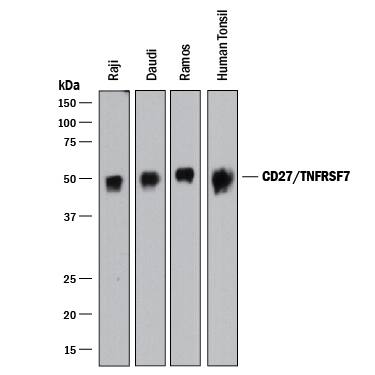
Detection of Human CD27/TNFRSF7 by Western Blot.
Western blot shows lysates of Raji human Burkitt's lymphoma cell line, Daudi human Burkitt's lymphoma cell line, Ramos human Burkitt's lymphoma cell line, and human tonsil tissue. PVDF membrane was probed with 1 µg/mL of Goat Anti-Human CD27/TNFRSF7 Antigen Affinity-purified Polyclonal Antibody (Catalog # AF382) followed by HRP-conjugated Anti-Goat IgG Secondary Antibody (HAF017). A specific band was detected for CD27/ TNFRSF7 at approximately 50 kDa (as indicated). This experiment was conducted under reducing conditions and using Immunoblot Buffer Group 1.CD27/TNFRSF7 in Human Tonsil.
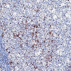
CD27/TNFRSF7 was detected in immersion fixed paraffin-embedded sections of human tonsil using Goat Anti-Human CD27/TNFRSF7 Antigen Affinity-purified Polyclonal Antibody (Catalog # AF382) at 3 µg/mL overnight at 4 °C. Tissue was stained using the Anti-Goat HRP-DAB Cell & Tissue Staining Kit (brown; CTS008) and counterstained with hematoxylin (blue). Specific staining was localized to lymphocytes in germinal center. View our protocol for Chromogenic IHC Staining of Paraffin-embedded Tissue Sections.Detection of Human CD27/TNFRSF7 by Simple WesternTM.
Simple Western lane view shows lysates of human tonsil tissue, loaded at 0.2 mg/mL. A specific band was detected for CD27/TNFRSF7 at approximately 49 kDa (as indicated) using 20 µg/mL of Goat Anti-Human CD27/ TNFRSF7 Antigen Affinity-purified Polyclonal Antibody (Catalog # AF382) followed by 1:50 dilution of HRP-conjugated Anti-Goat IgG Secondary Antibody (Catalog # HAF109). This experiment was conducted under reducing conditions and using the 12-230 kDa separation system.Applications for Human CD27/TNFRSF7 Antibody
CyTOF-ready
Flow Cytometry
Sample: Human whole blood lymphocytes
Immunohistochemistry
Sample: Immersion fixed paraffin-embedded sections of human tonsil
Simple Western
Sample: Human tonsil tissue
Western Blot
Sample: Raji human Burkitt's lymphoma cell line, Daudi human Burkitt's lymphoma cell line, Ramos human Burkitt's lymphoma cell line, and human tonsil tissue
Neutralization
Reviewed Applications
Read 1 review rated 5 using AF382 in the following applications:
Formulation, Preparation, and Storage
Purification
Reconstitution
Formulation
Shipping
Stability & Storage
- 12 months from date of receipt, -20 to -70 °C as supplied.
- 1 month, 2 to 8 °C under sterile conditions after reconstitution.
- 6 months, -20 to -70 °C under sterile conditions after reconstitution.
Background: CD27/TNFRSF7
Human CD27 is a lymphocyte-specific member of the TNF receptor superfamily. CD27 is expressed on a subset of human thymocytes and on the majority of mature T cells. CD27 expression is up-regulated after TCR stimulation. Within the CD4+ compartment, it is preferentially expressed on CD45RA+ cells. In contrast, it is preferentially expressed on CD45RO+ cells in the CD8+ compartment. CD27 also appeaars to be a potential marker for memory B cells. It exists as both a disulfide-linked dimer on the cell surface and as a soluble protein found in serum. Human CD27 is a 260 amino acid (aa) protein with a 20 aa signal, a 173 aa extracellular domain, a 20 aa transmembrane domain, and a 47 aa cytoplasmic domain. The ligand for CD27 is CD70. CD70 is expressed on thymic stromal cells and a small subset of activated T cells. Additionally a subset of activated B cells express CD70. The CD27/CD70 interaction appears to be a weak costimulatory pathway involved in T cell and B cell immune response. CD27/CD70 interactions may be more involved in controlling the expansion phase of an immune response. This would be in contrast to B7/CD28 interactions, which are important for the activation phase of immune responses.
References
- Camerini, D. et al. (1991) J. Immunol. 147:3165.
- Loenen, W.A. et al. (1992) J. Immunol. 149:3937.
- Lens, S.M.A. et al. (1998) Sem. Immunol. 10:491.
Alternate Names
Gene Symbol
UniProt
Additional CD27/TNFRSF7 Products
Product Documents for Human CD27/TNFRSF7 Antibody
Product Specific Notices for Human CD27/TNFRSF7 Antibody
For research use only


