Human CD36/SR-B3 Antibody
R&D Systems, part of Bio-Techne | Catalog # MAB19554

Key Product Details
Species Reactivity
Applications
Label
Antibody Source
Product Specifications
Immunogen
Accession # P16671
Specificity
Clonality
Host
Isotype
Scientific Data Images for Human CD36/SR-B3 Antibody
Detection of Human CD36/SR‑B3 by Western Blot.
Western blot shows lysates of human placenta tissue and human platelets. PVDF membrane was probed with 1 µg/mL of Rabbit Anti-Human CD36/SR-B3 Monoclonal Antibody (Catalog # MAB19554) followed by HRP-conjugated Anti-Rabbit IgG Secondary Antibody (Catalog # HAF008). A specific band was detected for CD36/SR-B3 at approximately 85 kDa (as indicated). This experiment was conducted under reducing conditions and using Immunoblot Buffer Group 1.CD36/SR‑B3 in U937 Human Cell Line.
CD36/SR-B3 was detected in immersion fixed U937 human histiocytic lymphoma cell line using Rabbit Anti-Human CD36/SR-B3 Monoclonal Antibody (Catalog # MAB19554) at 8 µg/mL for 3 hours at room temperature. Cells were stained using the NorthernLights™ 557-conjugated Anti-Rabbit IgG Secondary Antibody (red; Catalog # NL004) and counterstained with DAPI (blue). Specific staining was localized to cytoplasm. View our protocol for Fluorescent ICC Staining of Cells on Coverslips.CD36/SR‑B3 in Human Heart.
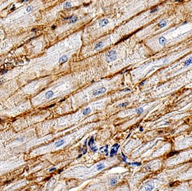
CD36/SR-B3 was detected in immersion fixed paraffin-embedded sections of human heart using Rabbit Anti-Human CD36/SR-B3 Monoclonal Antibody (Catalog # MAB19554) at 3 µg/mL for 1 hour at room temperature followed by incubation with the Anti-Rabbit IgG VisUCyte™ HRP Polymer Antibody (Catalog # VC003). Tissue was stained using DAB (brown) and counterstained with hematoxylin (blue). Specific staining was localized to cardiomyocyte membranes. View our protocol for IHC Staining with VisUCyte HRP Polymer Detection Reagents.Applications for Human CD36/SR-B3 Antibody
Immunocytochemistry
Sample: Immersion fixed U937 human histiocytic lymphoma cell line
Immunohistochemistry
Sample: Immersion fixed paraffin-embedded sections of human heart
Western Blot
Sample: Human placenta tissue and human platelets
Formulation, Preparation, and Storage
Purification
Reconstitution
Formulation
Shipping
Stability & Storage
- 12 months from date of receipt, -20 to -70 °C as supplied.
- 1 month, 2 to 8 °C under sterile conditions after reconstitution.
- 6 months, -20 to -70 °C under sterile conditions after reconstitution.
Background: CD36/SR-B3
CD36, alternatively known as platelet membrane glycoprotein IV (GPIV), GPIIIb, thrombospondin receptor, collagen receptor, fatty acid translocase (FAT), and scavenger receptor class B, member 3 (SR-B3), is an integral membrane glycoprotein that has multiple physiological functions (1). It is broadly expressed on a variety of cell types including microvascular endothelium, adipocytes, skeletal muscle, epithelial cells of the retina, breast, and intestine, smooth muscle cells, erythroid precursors, platelets, megakaryocytes, dendritic cells, monocytes/macrophages, and microglia (1, 2). As a member of the scavenger receptor family, CD36 is a multiligand pattern recognition receptor that interacts with a large number of structurally dissimilar ligands, including long chain fatty acid (LCFA), advanced glycation end products (AGE), thrombospondin-1, oxidized low-density lipoproteins (oxLDLs), high density lipoprotein (HDL), phosphatidylserine, apoptotic cells,
beta‑amyloid fibrils (fA beta), collagens I and IV, and Plasmodium falciparum-infected erythrocytes (3). CD36 is required for the anti-angiogenic effects of thrombospondin-1 in the corneal neovascularization assay (4). It plays a role in lipid metabolism and has been identified as a fatty acid translocase necessary for the binding and transport of LCFA in cells and tissues (5). CD36 has been implicated in the clearance of apoptotic cells and cell debris and has also been shown to mediate the internalization and degradation of a variety of its ligands such as oxLDL, AGE and fA beta (3). Upon ligand binding, CD36 transduces signals that mediate a wide range of pro-inflammatory cellular responses (2). CD36 plays a significant role in the initiation and pathogenesis of chronic inflammatory diseases such as Alzheimer’s disease and atherosclerosis (2, 3). The human CD36 gene encodes a single-chain 472 amino acid protein containing both an N- and a C-terminal cytoplasmic tail and an extracellular loop.
References
- Febbraio, M. et al. (2001) J. Clin. Invest. 108:785.
- Khoury, J. et al. (2003) J. Exp. Med. 197:1657.
- Husemann, J. et al. (2002) Glia 40:195.
- Armstrong, L and P. Bornstein (2003) Matrix. Biol. 22:63.
- Febbraio M. et al. (1999) J. Biol. Chem. 274:19055.
Long Name
Alternate Names
Gene Symbol
UniProt
Additional CD36/SR-B3 Products
Product Documents for Human CD36/SR-B3 Antibody
Product Specific Notices for Human CD36/SR-B3 Antibody
For research use only


