Human CD4 Antibody
R&D Systems, part of Bio-Techne | Catalog # MAB11560

Key Product Details
Species Reactivity
Applications
Label
Antibody Source
Product Specifications
Immunogen
Lys26-Trp390
Accession # P01730
Specificity
Clonality
Host
Isotype
Scientific Data Images for Human CD4 Antibody
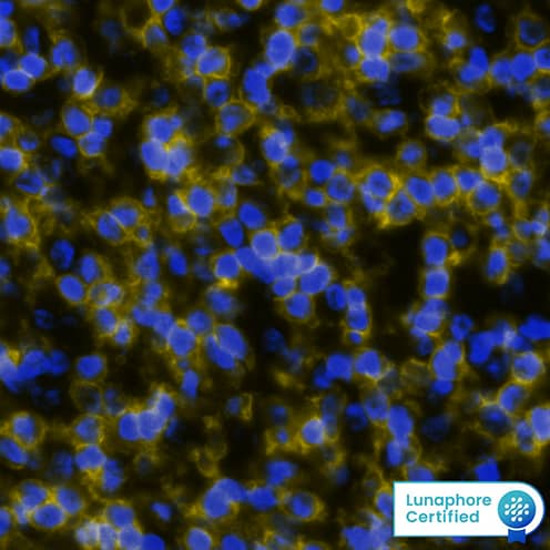
Detection of CD4 in Human Tonsil via Multiplex Immunofluorescence staining on COMET™
CD4 was detected in immersion fixed paraffin-embedded sections of human tonsil using Rabbit Anti-Human CD4 Monoclonal Antibody (Catalog # MAB11560) at 20µg/mL at 37 ° Celsius for 8 minutes. Before incubation with the primary antibody, tissue underwent an all-in-one dewaxing and antigen retrieval preprocessing using PreTreatment Module (PT Module) and Dewax and HIER Buffer H (pH 9). Tissue was stained using the Alexa Fluor™ Plus 647 Goat anti-Rabbit IgG Secondary Antibody at 1:200 at 37 ° Celsius for 2 minutes. (Yellow; Lunaphore Catalog # DR647RB) and counterstained with DAPI (blue; Lunaphore Catalog # DR100).Specific staining was localized to the membrane. Protocol available in COMET™ Panel Builder.Detection of CD4 in Human Tonsil.
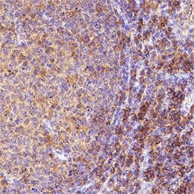
CD4 was detected in immersion fixed paraffin-embedded sections of human tonsil using Rabbit Anti-Human CD4 Monoclonal Antibody (Catalog # MAB11560) at 1 µg/ml for 1 hour at room temperature followed by incubation with the Anti-Rabbit IgG VisUCyte™ HRP Polymer Antibody (Catalog # VC003) or the HRP-conjugated Anti-Rabbit IgG Secondary Antibody (Catalog # HAF008). Before incubation with the primary antibody, tissue was subjected to heat-induced epitope retrieval using VisUCyte Antigen Retrieval Reagent-Basic (Catalog # VCTS021). Tissue was stained using DAB (brown) and counterstained with hematoxylin (blue). Specific staining was localized to the cytoplasm and membrane. View our protocol for Chromogenic IHC Staining of Paraffin-embedded Tissue Sections.Detection of CD4 in hPBMC cells (Positive) and MCF-7 cells (Negative).
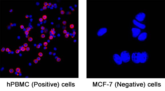
CD4 was detected in fixed hPBMC cells (Positive) and absent in MCF-7 human breast cancer cell line (Negative) using Rabbit Anti-Human CD4 Monoclonal Antibody (Catalog # MAB11560) at 3 µg/ml for 3 hours at room temperature. Cells were stained using the NorthernLights™ 557-conjugated Anti-Rabbit IgG Secondary Antibody (red; Catalog # NL004) and counterstained with DAPI (blue). Specific staining was localized to the membrane. View our protocol for Fluorescent ICC Staining of Non-adherent Cells.Applications for Human CD4 Antibody
Immunocytochemistry
Sample: fixed hPBMC cells (Positive) and absent in MCF-7 human breast cancer cell line (Negative)
Immunohistochemistry
Sample: Immersion fixed paraffin-embedded sections of human tonsil
Multiplex Immunofluorescence
Sample: Immersion fixed paraffin embedded sections of human tonsil
Formulation, Preparation, and Storage
Purification
Reconstitution
Formulation
Shipping
Stability & Storage
- 12 months from date of receipt, -20 to -70 °C as supplied.
- 1 month, 2 to 8 °C under sterile conditions after reconstitution.
- 6 months, -20 to -70 °C under sterile conditions after reconstitution.
Background: CD4
CD4 is an approximately 55 kDa type I membrane glycoprotein that is expressed predominantly on most thymocytes and a subset of mature T lymphocytes. In humans, CD4 is also expressed to a lesser extent on monocytes and macrophage related cells. Human CD4 cDNA encodes a 458 amino acid (aa) precursor protein with a 25 aa signal peptide, a 371 aa extracellular region containing four immunoglobulin homology domains, a 24 aa transmembrane domain and a 38 aa cytoplasmic domain. CD4 is a coreceptor required for T cell recognition of antigens that are presented by class II major histocompatibility complexes. CD4 has been shown to be a coreceptor of HIV entry and specifically binds gp120, the external envelope glycoprotein of HIV.
References
- Capon, D.I. et al. (1991) Annu. Rev. Immunol. 9:649.
Alternate Names
Entrez Gene IDs
Gene Symbol
UniProt
Additional CD4 Products
Product Documents for Human CD4 Antibody
Product Specific Notices for Human CD4 Antibody
For research use only


