Human CD99 Antibody
R&D Systems, part of Bio-Techne | Catalog # MAB3968


Key Product Details
Species Reactivity
Applications
Label
Antibody Source
Product Specifications
Immunogen
Asp23-Asp122
Accession # P14209
Specificity
Clonality
Host
Isotype
Scientific Data Images for Human CD99 Antibody
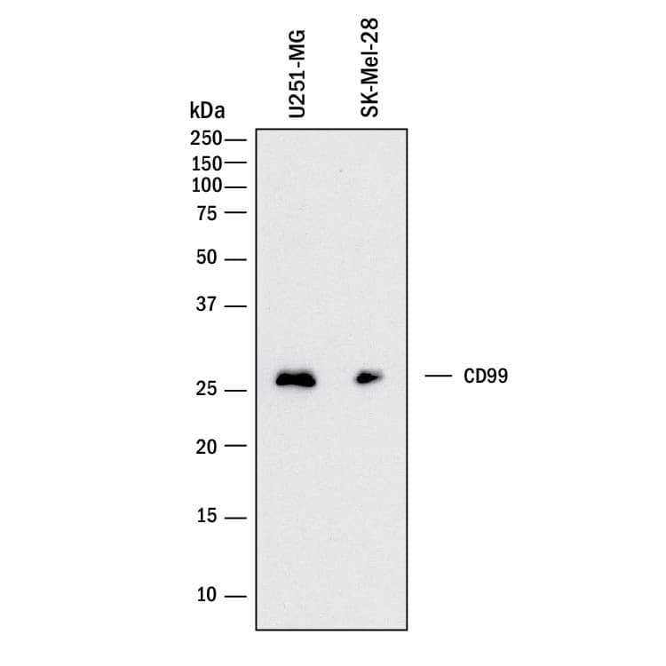
Detection of Human CD99 by Western Blot.
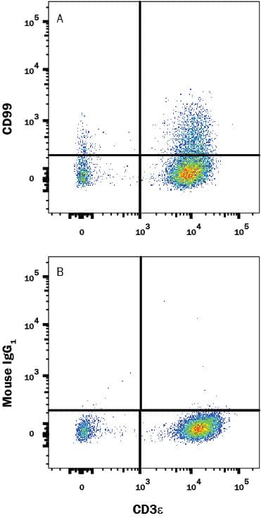
Western blot shows lysates of U251-MG human malignant glioblastoma cell line and SK-Mel-28 human malignant melanoma cell line. PVDF membrane was probed with 2 µg/mL of Mouse Anti-Human CD99 Monoclonal Antibody (Catalog # MAB3968) followed by HRP-conjugated Anti-Mouse IgG Secondary Antibody (Catalog # HAF018). A specific band was detected for CD99 at approximately 30 kDa (as indicated). This experiment was conducted under reducing conditions and using Immunoblot Buffer Group 1.Detection of CD99 in Human PBMC Lymphocytes by Flow Cytometry.
Human peripheral blood mononuclear cell (PBMC) lymphocytes were stained with (A) Mouse Anti-Human CD99 Monoclonal Antibody (Catalog # MAB3968) or (B) Mouse IgG1 Isotype Control (Catalog # MAB002) followed by anti-Mouse IgG PE-conjugated secondary antibody (Catalog # F0102B) and Mouse Anti-Human CD3 APC-conjugated Monoclonal Antibody (Catalog # FAB100A). View our protocol for Staining Membrane-associated Proteins.Applications for Human CD99 Antibody
CyTOF-ready
Flow Cytometry
Sample: Human PBMC lymphocytes
Western Blot
Sample: U251-MG human malignant glioblastoma cell line and SK‑Mel‑28 human malignant melanoma cell line
Formulation, Preparation, and Storage
Purification
Reconstitution
Formulation
Shipping
Stability & Storage
- 12 months from date of receipt, -20 to -70 °C as supplied.
- 1 month, 2 to 8 °C under sterile conditions after reconstitution.
- 6 months, -20 to -70 °C under sterile conditions after reconstitution.
Background: CD99
CD99 (also named MIC2, E2 and thymic leukemia antigen) is the founding member of the CD99 family of molecules. The CD99 family contains four members; CD99, CD99L2, XG and the pseudogene CD99L1 (1, 2, 3). Native human CD99 is 32 kDa in size and exists as a type I transmembrane glycoprotein. This is referred to as the long, or type I isoform. It is synthesized as a 185 amino acid (aa) precursor that contains a 22 aa signal sequence, a 100 aa extracellular domain (ECD), a 25 aa transmembrane segment, and a 38 aa cytoplasmic region (4). The ECD contains no identifiable motifs, N-linked glycosylation sites, or cysteine residues; it does possess sites for O-linked glycosylation. The cytoplasmic region, albeit short, does have signal transduction capability (5). There are apparently multiple isoforms for human CD99. One shows a 16 aa deletion in the ECD (aa 34 - 49), a second shows a 38 aa deletion in the cytoplasmic region (aa 122 - 159), and a third exhibits a three aa truncation at the C-terminus (6, 7, 8). The best studied isoform shows an Asp-Gly substitution for the C-terminal 27 amino acids. This is referred to as the 28 kDa type II isoform (9). The type I and II isoforms have distinctive signal transduction pathways (FAK-src for type I; PI3K plus src-ERK1/2 for type II), and mediate clearly different biological outcomes (5, 9, 10). The two numbered isoforms may or may not coexist on the same cells. Peripheral T cells have only the long isoform, while double-positive thymocytes express both isotypes. What is unclear is the monomeric vs. dimeric status of CD99. In mouse, CD99 reportedly forms disulfide-linked homodimers (11). In human, however, CD99 is reportedly monomeric if only a type I isoform, and a covalent heterodimer if coexpressing type I and II isoforms (12, 13). Cells known to express CD99 include fibroblasts, neutrophils, T cells, double-positive thymocytes, CD34+ stem cells, monocytes and endothelial cells (2, 12, 14, 15). Homophilic interaction between CD99 on the neutrophil and CD99 on the endothelial cell regulates the transendothelial migration of neutrophils during inflammation (16). Human CD99 is only 48% aa identical to mouse CD99 (17).
References
- Wilson, M.D. et al. (2006) Physiol Genomics 27:201.
- Petri, B. and M.G. Bixel (2006) FEBS J. 273:4399.
- Suh, Y.H. et al. (2003) Gene 307:63.
- Gelin, C. et al. (1989) EMBO J. 8:3253.
- Byun, H-J. et al. (2006) J. Biol. Chem. 281:34833.
- GenBank Accession # EAW98698.
- GenBank Accession # EAW98699.
- GenBank Accession # EAW98700.
- Hahn, H-J. et al. (1997) J. Immunol. 159:2250.
- Scotlandi, K. et al. (2007) Oncogene Apr 30; [Epub ahead of print].
- Park, S.H. et al. (2005) Gene 353:177.
- Schenkel, A.R. et al. (2002) Nat. Immunol. 3:143.
- Alberti, I. et al. (2002) FASEB J. 16:1946.
- Imbert, A-M. et al. (2006) Blood 108:2578.
- Dworzak, M.N. et al. (1994) Blood 83:415.
- Lou, O. et al. (2007) J. Immunol. 178:1136
- Shiratori, I. et al. (2004) J. Exp. Med. 199:525.
Alternate Names
Gene Symbol
UniProt
Additional CD99 Products
Product Documents for Human CD99 Antibody
Product Specific Notices for Human CD99 Antibody
For research use only
