Human CD99-L2 Antibody
R&D Systems, part of Bio-Techne | Catalog # AF5185

Key Product Details
Species Reactivity
Validated:
Cited:
Applications
Validated:
Cited:
Label
Antibody Source
Product Specifications
Immunogen
Val20-Ala188
Accession # Q8TCZ2
Specificity
Clonality
Host
Isotype
Scientific Data Images for Human CD99-L2 Antibody
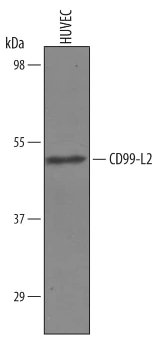
Detection of Human CD99‑L2 by Western Blot.
Western blot shows lysates of HUVEC human umbilical vein endothelial cells. PVDF Membrane was probed with 1 µg/mL of Sheep Anti-Human CD99-L2 Antigen Affinity-purified Polyclonal Antibody (Catalog # AF5185) followed by HRP-conjugated Anti-Sheep IgG Secondary Antibody (HAF016). A specific band was detected for CD99-L2 at approximately 50 kDa (as indicated). This experiment was conducted under reducing conditions and using Immunoblot Buffer Group 1.Applications for Human CD99-L2 Antibody
Western Blot
Sample: HUVEC human umbilical vein endothelial cells
Formulation, Preparation, and Storage
Purification
Reconstitution
Formulation
Shipping
Stability & Storage
- 12 months from date of receipt, -20 to -70 °C as supplied.
- 1 month, 2 to 8 °C under sterile conditions after reconstitution.
- 6 months, -20 to -70 °C under sterile conditions after reconstitution.
Background: CD99-L2
CD99 antigen-like 2 (CD99-L2) is a 45 kDa type I transmembrane glycoprotein in the CD99 family of molecules (1‑3). The major form of human CD99-L2 cDNA encodes a 262 amino acid (aa) precursor with a 25 aa predicted signal sequence, a 160 aa extracellular domain (ECD), a 21 aa transmembrane (TM) segment, and a 56 aa cytoplasmic region (4). This form is called the long form, or isoform E3’-E4’-E3-E4. Other isoforms include the muscle E3-E4, missing aa 45‑93, and E4, a short form missing aa 45‑116 (1, 4). E3-E4 and E3’-E3-E4 forms are the major isoforms in mouse and rat, respectively (1). Human forms that diverge at Pro 45 (145 aa precursor) or Met 180 (173 aa precursor) have been sequenced, and would be predicted to lack a TM segment (4, 5). None of the forms contain predicted N-linked glycosylation sites within the ECD, but O-linked glycosylation is likely (1, 2). The ECD of the human CD99-L2 isoform E3-E4 shares 85%, 75% and 70% aa identity with the corresponding forms of mouse, rat, and bovine CD99-L2, respectively. The human CD99 and CD99-L2 ECDs share only about 35% aa identity, but both contain three conserved acidic motifs and are thought to originate from the same ancestral gene (1, 2). The nearly ubiquitous expression of CD99-L2 is similar to that of CD99. Human CD99-L2 cDNA is detected in most organs, but not in thymus (1). In the mouse, protein is detectable in lung, thymocytes, mouse leukocytes and vascular endothelial cells (1, 3, 6). The endothelial cell CD99-L2 is reported to mediate cell aggregation and neutrophil or monocyte, but not lymphocyte, extravasation to inflamed tissue in vivo (3, 6).
References
- Suh, Y.H. et al. (2003) Gene 307:63.
- Park, S.H. et al. (2005) Gene 353:177.
- Schenkel, A.R. et al. (2007) Cell Commun. Adhes. 14:227.
- Swissprot Accession # Q8TCZ2.
- Entrez Accession # EAW99391.
- Bixel, G. et al. (2007) Blood 109:5327.
Long Name
Alternate Names
Gene Symbol
UniProt
Additional CD99-L2 Products
Product Documents for Human CD99-L2 Antibody
Product Specific Notices for Human CD99-L2 Antibody
For research use only
