Human CHL-1/L1CAM-2 Antibody
R&D Systems, part of Bio-Techne | Catalog # MAB2126

Key Product Details
Validated by
Species Reactivity
Validated:
Cited:
Applications
Validated:
Cited:
Label
Antibody Source
Product Specifications
Immunogen
Ile25-Gln1096
Accession # NP_006605
Specificity
Clonality
Host
Isotype
Scientific Data Images for Human CHL-1/L1CAM-2 Antibody
CHL-1/L1CAM-2 in Human Melanoma Tissue.
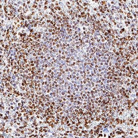
CHL-1/L1CAM-2 was detected in immersion fixed paraffin-embedded sections of human melanoma tissue using Rat Anti-Human CHL-1/L1CAM-2 Monoclonal Antibody (Catalog # MAB2126) at 1.7 µg/mL for 1 hour at room temperature followed by incubation with the Anti-Rat IgG VisUCyte™ HRP Polymer Antibody (VC005). Before incubation with the primary antibody, tissue was subjected to heat-induced epitope retrieval using Antigen Retrieval Reagent-Basic (CTS013). Tissue was stained using DAB (brown) and counterstained with hematoxylin (blue). Specific staining was localized to cell nuclei. Staining was performed using our protocol for IHC Staining with VisUCyte HRP Polymer Detection Reagents.CHL-1/L1CAM-2 in Human Spleen.
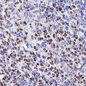
CHL-1/L1CAM-2 was detected in immersion fixed paraffin-embedded sections of human spleen using Rat Anti-Human CHL-1/L1CAM-2 Monoclonal Antibody (Catalog # MAB2126) at 1.7 µg/mL for 1 hour at room temperature followed by incubation with the Anti-Rat IgG VisUCyte™ HRP Polymer Antibody (VC005). Before incubation with the primary antibody, tissue was subjected to heat-induced epitope retrieval using Antigen Retrieval Reagent-Basic (CTS013). Tissue was stained using DAB (brown) and counterstained with hematoxylin (blue). Specific staining was localized to cell nuclei. Staining was performed using our protocol for IHC Staining with VisUCyte HRP Polymer Detection Reagents.Detection of Human CHL-1/L1CAM-2 by Western Blot
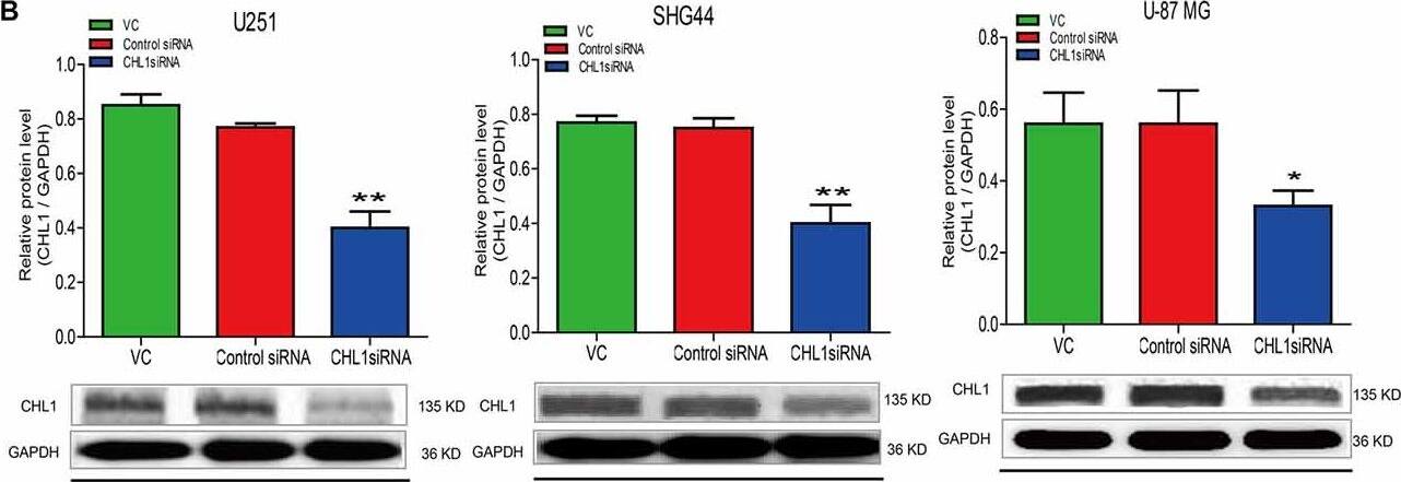
Western blot analysis of the protein levels of CHL1 detected in normal human glial HEB cells and 3 glioma/glioblastoma cell lines. CHL1 was weakly expressed in normal human HEB glial cells. Its levels in all the 3 glioma/glioblastoma cells were higher than that in normal human HEB glial cells, with the statistical significance detected in SHG44 cells (*p < 0.05 vs. HEB cells) and U-87 MG cells (**p < 0.01 vs. HEB cells). n = 3 for each group. Student’s t-test for independent samples was used. Image collected and cropped by CiteAb from the following publication (https://journal.frontiersin.org/article/10.3389/fnmol.2017.00324/full), licensed under a CC-BY license. Not internally tested by R&D Systems.Applications for Human CHL-1/L1CAM-2 Antibody
Immunohistochemistry
Sample: Immersion fixed paraffin-embedded sections of human melanoma tissue and human spleen
Western Blot
Sample: Recombinant Human CHL-1/L1CAM-2 (Catalog # 2126-CH)
Reviewed Applications
Read 1 review rated 5 using MAB2126 in the following applications:
Formulation, Preparation, and Storage
Purification
Reconstitution
Formulation
Shipping
Stability & Storage
- 12 months from date of receipt, -20 to -70 °C as supplied.
- 1 month, 2 to 8 °C under sterile conditions after reconstitution.
- 6 months, -20 to -70 °C under sterile conditions after reconstitution.
Background: CHL-1/L1CAM-2
Close homolog of L1 (CHL-1), also known as cell adhesion L1-like (CALL) and L1 cell adhesion molecule 2 (L1-CAM2), belongs to the L1 subfamily of the Ig superfamily cell adhesion molecules, which also include L1, neurofascin and NgCAM-related cell adhesion molecule (NrCAM) (1‑3). These molecules are type I transmembrane proteins that have 6 Ig-like domains and 4‑5 fibronectin type III-like (FNIII) domains in their extracellular regions. They also shared a highly conserved cytoplasmic region of approximately 110 amino acids (aa) containing an ankyrin-binding site. CHL-1 is expressed as a highly glycosylated 185 kDa transmembrane protein by subpopulations of neurons and glia of the central and peripheral nervous system (4, 5). Ectodomain shedding via the metalloprotease-disintegrin ADAM8 releases 165 kDa and 125 kDa soluble CHL-1 fragments, which can diffuse away to function at distant sites (6). CHL-1 is not capable of homotypic interactions, but an extracellular binding partner of CHL-1 has not been identified (4). Human CHL1 has been mapped to chromosome 3p26 and is a candidate gene for 3p- syndrome characterized by mental impairment (7). A missense CHL1 polymorphism associated with an increased risk of schizophrenia has been reported (8). The functional importance of CHL-1 in the nervous system is also evident in CHL-1 deficient mice, which display behavioral abnormalities and show misguided axons within the hippocampus and olfactory tract (9). Enhanced ectodomain-shedding of CHL-1 is also observed in Wobbler mice, the neurodegenerative mutant mice (6). In vitro, soluble or substrate-coated CHL-1 promotes neurite outgrowth and neuronal survival of both cerebellar and hippocampal neurons. Cell surface CHL-1 interacts with integrins in cis to potentiate integrin-dependent cell migration toward extracellular matrix proteins (10). For this enhanced cell motility, CHL-1 linkage to the actin cytoskeleton via interaction between ankyrin and the CHL-1 cytoplasmic region is required.
References
- Moos, M. et al. (1988) Nature 334:701.
- Holm, J. et al. (1996) Eur. J. Neusci. 8:1613.
- Wei, M. et al. (1998) Hum. Genet. 103:355.
- Hillenbrand, R. et al. (1999) Eur. J. Neurosci. 11:813.
- Liu, Q. et al. (2000) J. Neurosci. 20:7682.
- Naus, S. et al. (2004) J. Biol. Chem. 279:16083.
- Angeloni, D. et al. (1999) Am. J. Med. Genet. 86:482.
- Sakurai, K. et al. (2002) Mol. Psychiatry 7:412.
- Montag-Sallaz, M. et al. (2002) Mol. Cell. Biol. 22(22):7967.
- Buhusi, M. et al. (2003) J. Biol. Chem. 278(27):25024.
Long Name
Alternate Names
Gene Symbol
UniProt
Additional CHL-1/L1CAM-2 Products
Product Documents for Human CHL-1/L1CAM-2 Antibody
Product Specific Notices for Human CHL-1/L1CAM-2 Antibody
For research use only


