Human cIAP-1/HIAP-2 Antibody
R&D Systems, part of Bio-Techne | Catalog # MAB8181

Key Product Details
Validated by
Knockout/Knockdown
Species Reactivity
Human
Applications
Immunohistochemistry
Label
Unconjugated
Antibody Source
Monoclonal Mouse IgG1 Clone # 681724
Product Specifications
Immunogen
E. coli-derived recombinant human cIAP-1/HIAP-2
His2-Ser618
Accession # Q13490
His2-Ser618
Accession # Q13490
Specificity
Detects human cIAP-1/HIAP-2 in direct ELISAs. In direct ELISAs, 25% cross-reactivity with recombinant human cIAP-2 is observed.
Clonality
Monoclonal
Host
Mouse
Isotype
IgG1
Scientific Data Images for Human cIAP-1/HIAP-2 Antibody
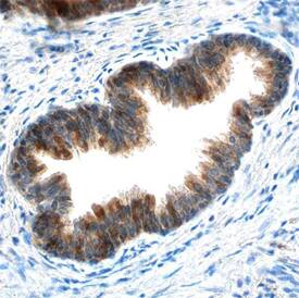
cIAP‑1/HIAP‑2 in Human Prostate.
cIAP-1/HIAP-2 was detected in immersion fixed paraffin-embedded sections of human prostate using Mouse Anti-Human cIAP-1/HIAP-2 Monoclonal Antibody (Catalog # MAB8181) at 15 µg/mL overnight at 4 °C. Before incubation with the primary antibody, tissue was subjected to heat-induced epitope retrieval using Antigen Retrieval Reagent-Basic (Catalog # CTS013). Tissue was stained using the Anti-Mouse HRP-DAB Cell & Tissue Staining Kit (brown; Catalog # CTS002) and counterstained with hematoxylin (blue). Specific staining was localized to the cytoplasm of epithelial cells. View our protocol for Chromogenic IHC Staining of Paraffin-embedded Tissue Sections.Detection of Human cIAP-1/HIAP-2 by Knockdown Validated
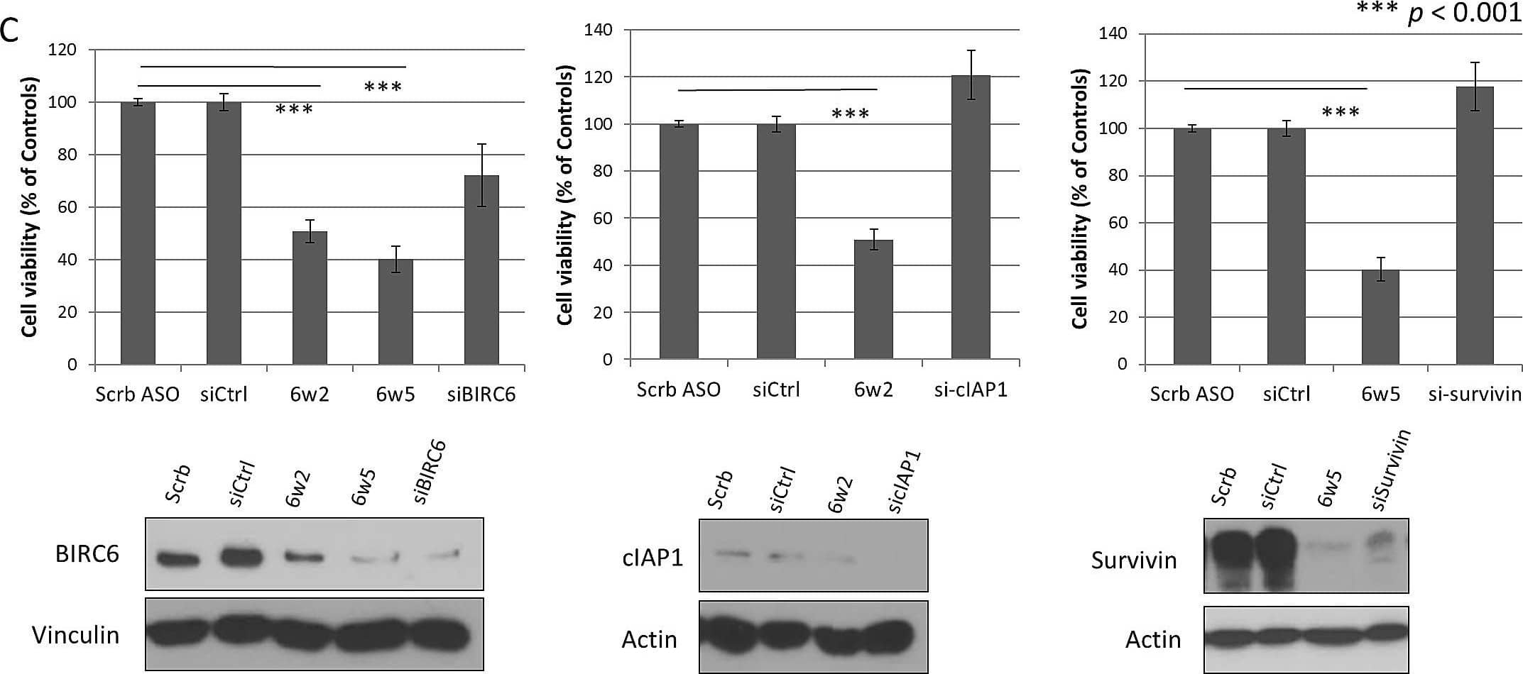
Dual IAP-targeting ASOs knockdown BIRC6, cIAP1 or survivin proteins and lead to marked suppression of CRPC cell proliferation(A-B) Western blotting showing protein levels of BIRC6, cIAP1 and survivin in two CRPC cell lines (A) PC-3 cells and (B) C4-2 cells transfected with Mock or increasing dosages of scrambled ASO (Scrb), dASOs 6w2 and 6w5 for 72 hr. (C) Comparison of dual IAP targeting and single IAP-targeting. Cell viability of PC-3 cells transfected with dASOs 6w2, 6w5 and siRNA-targeting BIRC6, cIAP1 or survivin, was determined by MTS assay at 72 hr after transfection. Cell viabilities of ASO- and siRNA-treated cells were normalized with corresponding Scrb ASO and siRNA controls. Error bars represent mean percentage cell viability ± S.D. Western blotting of 3 IAPs showing comparable amounts of reduced protein expression obtained with dASO and siRNA single IAP-targeting. (D) Proliferation of PC-3 cells transfected with mock, Scrb ASO, dASOs 6w2 and 6w5. Error bars represent mean cell number ± S.D. (E) MTS viability assay of C4-2 cells treated with dASOs. Image collected and cropped by CiteAb from the following publication (https://pubmed.ncbi.nlm.nih.gov/25071009), licensed under a CC-BY license. Not internally tested by R&D Systems.Applications for Human cIAP-1/HIAP-2 Antibody
Application
Recommended Usage
Immunohistochemistry
8-25 µg/mL
Sample: Immersion fixed paraffin-embedded sections of human prostate
Sample: Immersion fixed paraffin-embedded sections of human prostate
Formulation, Preparation, and Storage
Purification
Protein A or G purified from hybridoma culture supernatant
Reconstitution
Sterile PBS to a final concentration of 0.5 mg/mL. For liquid material, refer to CoA for concentration.
Formulation
Lyophilized from a 0.2 μm filtered solution in PBS with Trehalose. *Small pack size (SP) is supplied either lyophilized or as a 0.2 µm filtered solution in PBS.
Shipping
Lyophilized product is shipped at ambient temperature. Liquid small pack size (-SP) is shipped with polar packs. Upon receipt, store immediately at the temperature recommended below.
Stability & Storage
Use a manual defrost freezer and avoid repeated freeze-thaw cycles.
- 12 months from date of receipt, -20 to -70 °C as supplied.
- 1 month, 2 to 8 °C under sterile conditions after reconstitution.
- 6 months, -20 to -70 °C under sterile conditions after reconstitution.
Background: cIAP-1/HIAP-2
References
- Roy, N. et al. (1997) EMBO J. 23:6914.
- Deveraux, Q. et al. (1997) Nature 388:300.
- Deveraux, Q. and J. Reed (1999) Genes & Develop. 13:239.
- Herrera, B. et al. (2002) FEBS Letters 520:93.
Long Name
Cellular Inhibitor of Apoptosis Protein 1
Alternate Names
BIRC2, cIAP1, HIAP-2, MIHB
Gene Symbol
BIRC2
UniProt
Additional cIAP-1/HIAP-2 Products
Product Documents for Human cIAP-1/HIAP-2 Antibody
Product Specific Notices for Human cIAP-1/HIAP-2 Antibody
For research use only
Loading...
Loading...
Loading...
Loading...

