Human CMG-2/ANTXR2 Antibody
R&D Systems, part of Bio-Techne | Catalog # AF2940


Key Product Details
Validated by
Biological Validation
Species Reactivity
Validated:
Human
Cited:
Human
Applications
Validated:
CyTOF-ready, ELISA, Flow Cytometry, Western Blot
Cited:
Flow Cytometry, Immunoprecipitation, STED Microscopy, Western Blot
Label
Unconjugated
Antibody Source
Polyclonal Goat IgG
Product Specifications
Immunogen
Mouse myeloma cell line NS0-derived recombinant human CMG‑2/ANTXR2 isoform 1
Gln34-Asn317
Accession # P58335
Gln34-Asn317
Accession # P58335
Specificity
Detects human CMG‑2/ANTXR2 in direct ELISAs and Western blots. In direct ELISAs and Western blots, approximately 20% cross-reactivity with recombinant mouse CMG-2 is observed.
Clonality
Polyclonal
Host
Goat
Isotype
IgG
Scientific Data Images for Human CMG-2/ANTXR2 Antibody
Detection of CMG-2/ANTXR2 in Human Monocytes by Flow Cytometry.
Human whole blood monocytes were stained with Human CMG-2/ANTXR2 Antigen Affinity-purified Polyclonal Antibody (Catalog # AF2940, filled histogram) or control antibody (Catalog # AB-108-C, open histogram), followed by Phycoerythrin-conjugated Anti-Goat IgG Secondary Antibody (Catalog # F0107).Detection of Human CMG-2/ANTXR2 by Western Blot
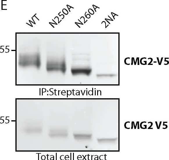
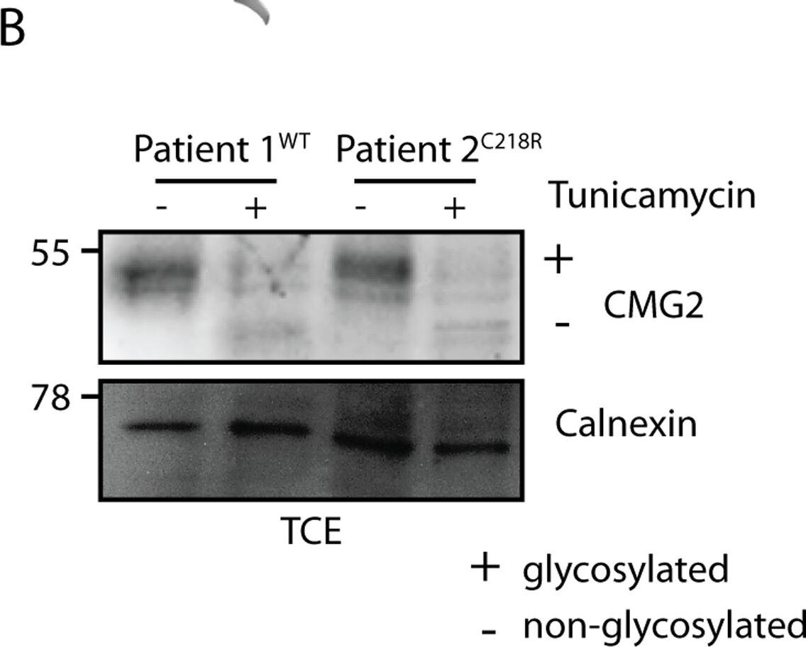
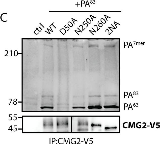
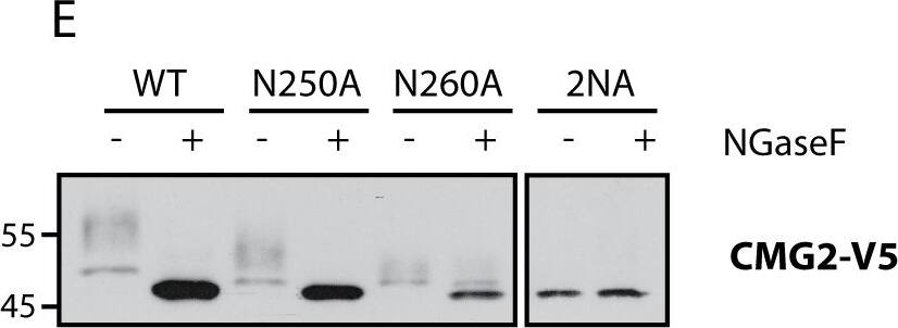
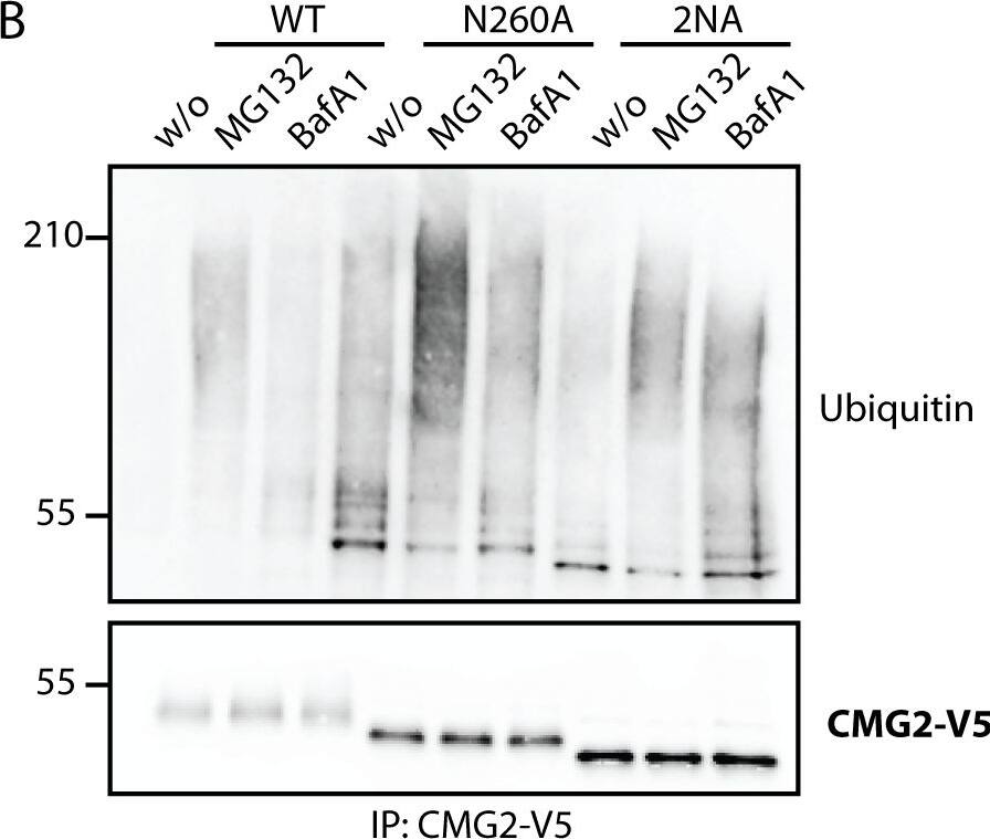
Number and localization of glycan sidechains determine trafficking efficiency of TEM8 and CMG2.A) Endoglycosidase H (EndoH) treatment on TEM8 and CMG2 single mutants. HeLa cells were transfected for 48h with the respective cDNAs. 40 μg of cell extracts were treated or not with EndoH as described before. Samples were analyzed by SDS-PAGE and Western Blotting. B) Quantification of surface biotinylation experiments to determine amount of TEM8 at the cell surface. All mutants were corrected for their expression levels and then normalized to WT, which was set at 100%. Errors represent standard deviation. Statistics were calculated using an unpaired t-test. n ≥ 3. * p≤0.05, ** p≤0.01, *** p≤0.001 C) Representative Western Blots of surface biotinylation. HeLa cells were transfected 48h with the respective cDNAs. Proteins at the cell surface were labeled with biotin, immunoprecipitated with streptavidin beads and blotted against TEM8-HA. D) Quantification of surface biotinylation experiments to determine amount of CMG2 at the cell surface. All mutants were corrected for their expression levels and then normalized to WT, which was set at 100%. Errors represent standard deviation. Statistics were calculated using an unpaired t-test. n ≥ 3. * p≤0.05, ** p≤0.01, *** p≤0.001 E) Representative Western Blots of surface biotinylation. HeLa cells were transfected 48h with the respective cDNAs. Proteins at the cell surface were labeled with biotin, immunoprecipitated with streptavidin beads and blotted against CMG2-V5. Image collected and cropped by CiteAb from the following publication (https://pubmed.ncbi.nlm.nih.gov/25781883), licensed under a CC-BY license. Not internally tested by R&D Systems.Detection of Human CMG-2/ANTXR2 by Western Blot
Number and localization of glycan sidechains determine trafficking efficiency of TEM8 and CMG2.A) Endoglycosidase H (EndoH) treatment on TEM8 and CMG2 single mutants. HeLa cells were transfected for 48h with the respective cDNAs. 40 μg of cell extracts were treated or not with EndoH as described before. Samples were analyzed by SDS-PAGE and Western Blotting. B) Quantification of surface biotinylation experiments to determine amount of TEM8 at the cell surface. All mutants were corrected for their expression levels and then normalized to WT, which was set at 100%. Errors represent standard deviation. Statistics were calculated using an unpaired t-test. n ≥ 3. * p≤0.05, ** p≤0.01, *** p≤0.001 C) Representative Western Blots of surface biotinylation. HeLa cells were transfected 48h with the respective cDNAs. Proteins at the cell surface were labeled with biotin, immunoprecipitated with streptavidin beads and blotted against TEM8-HA. D) Quantification of surface biotinylation experiments to determine amount of CMG2 at the cell surface. All mutants were corrected for their expression levels and then normalized to WT, which was set at 100%. Errors represent standard deviation. Statistics were calculated using an unpaired t-test. n ≥ 3. * p≤0.05, ** p≤0.01, *** p≤0.001 E) Representative Western Blots of surface biotinylation. HeLa cells were transfected 48h with the respective cDNAs. Proteins at the cell surface were labeled with biotin, immunoprecipitated with streptavidin beads and blotted against CMG2-V5. Image collected and cropped by CiteAb from the following publication (https://pubmed.ncbi.nlm.nih.gov/25781883), licensed under a CC-BY license. Not internally tested by R&D Systems.Applications for Human CMG-2/ANTXR2 Antibody
Application
Recommended Usage
CyTOF-ready
Ready to be labeled using established conjugation methods. No BSA or other carrier proteins that could interfere with conjugation.
ELISA
This antibody functions as an ELISA detection antibody when paired with Mouse Anti-Human CMG‑2/ANTXR2 Monoclonal Antibody (Catalog # MAB29401).
This product is intended for assay development on various assay platforms requiring antibody pairs.
Flow Cytometry
2.5 µg/106 cells
Sample: Human whole blood monocytes
Sample: Human whole blood monocytes
Western Blot
0.1 µg/mL
Sample: Recombinant Human CMG-2/ANTXR2 (Catalog # 2940-CM)
Sample: Recombinant Human CMG-2/ANTXR2 (Catalog # 2940-CM)
Formulation, Preparation, and Storage
Purification
Antigen Affinity-purified
Reconstitution
Reconstitute at 0.2 mg/mL in sterile PBS. For liquid material, refer to CoA for concentration.
Formulation
Lyophilized from a 0.2 μm filtered solution in PBS with Trehalose. *Small pack size (SP) is supplied either lyophilized or as a 0.2 µm filtered solution in PBS.
Shipping
Lyophilized product is shipped at ambient temperature. Liquid small pack size (-SP) is shipped with polar packs. Upon receipt, store immediately at the temperature recommended below.
Stability & Storage
Use a manual defrost freezer and avoid repeated freeze-thaw cycles.
- 12 months from date of receipt, -20 to -70 °C as supplied.
- 1 month, 2 to 8 °C under sterile conditions after reconstitution.
- 6 months, -20 to -70 °C under sterile conditions after reconstitution.
Background: CMG-2/ANTXR2
References
- Scobie, H.M. and J.A.T. Young (2005) Curr. Opin. Microbiol. 8:106.
- Scobie, H.M. et al. (2003) Proc. Natl. Acad. Sci. USA 100:5170.
- Bell, S.E. et al. (2001) J. Cell Sci. 114:2755.
- Lacy, D.B. et al. (2004) Proc. Natl. Acad. Sci. USA 101:6367.
- Santelli, E. et al. (2004) Nature 430:905.
- Dowling, O. et al. (2003) Am. J. Hum. Genet. 73:957.
- Wigelsworth, D.J. et al. (2004) J. Biol. Chem. 279:23349.
- Go, M.Y. et al. (2006) J. Mol. Biol. 360:145.
- Abrami, L. et al. (2006) J. Cell Biol. 172:309.
- Wei, W. et al. (2006) Cell 124:1141.
- Lacy, D.B. et al. (2004) Proc. Natl. Acad. Sci. USA 101:13147.
Long Name
Capillary Morphogenesis Protein 2
Alternate Names
ANTXR2, cI-35, CMG2, ISH, JHS
Gene Symbol
ANTXR2
UniProt
Additional CMG-2/ANTXR2 Products
Product Documents for Human CMG-2/ANTXR2 Antibody
Product Specific Notices for Human CMG-2/ANTXR2 Antibody
For research use only
Loading...
Loading...
Loading...
Loading...
Loading...

