Human CREG Antibody
R&D Systems, part of Bio-Techne | Catalog # MAB2380

Key Product Details
Species Reactivity
Validated:
Human
Cited:
Human
Applications
Validated:
Western Blot
Cited:
Immunohistochemistry-Frozen, Western Blot
Label
Unconjugated
Antibody Source
Monoclonal Mouse IgG2B Clone # 299133
Product Specifications
Immunogen
Mouse myeloma cell line NS0-derived recombinant human CREG
Arg32-Gln220
Accession # O75629
Arg32-Gln220
Accession # O75629
Specificity
Detects human CREG in direct ELISAs and Western blots. In direct ELISAs and Western blots, no cross-reactivity with recombinant mouse CREG is observed.
Clonality
Monoclonal
Host
Mouse
Isotype
IgG2B
Scientific Data Images for Human CREG Antibody
Detection of Human CREG by Western Blot
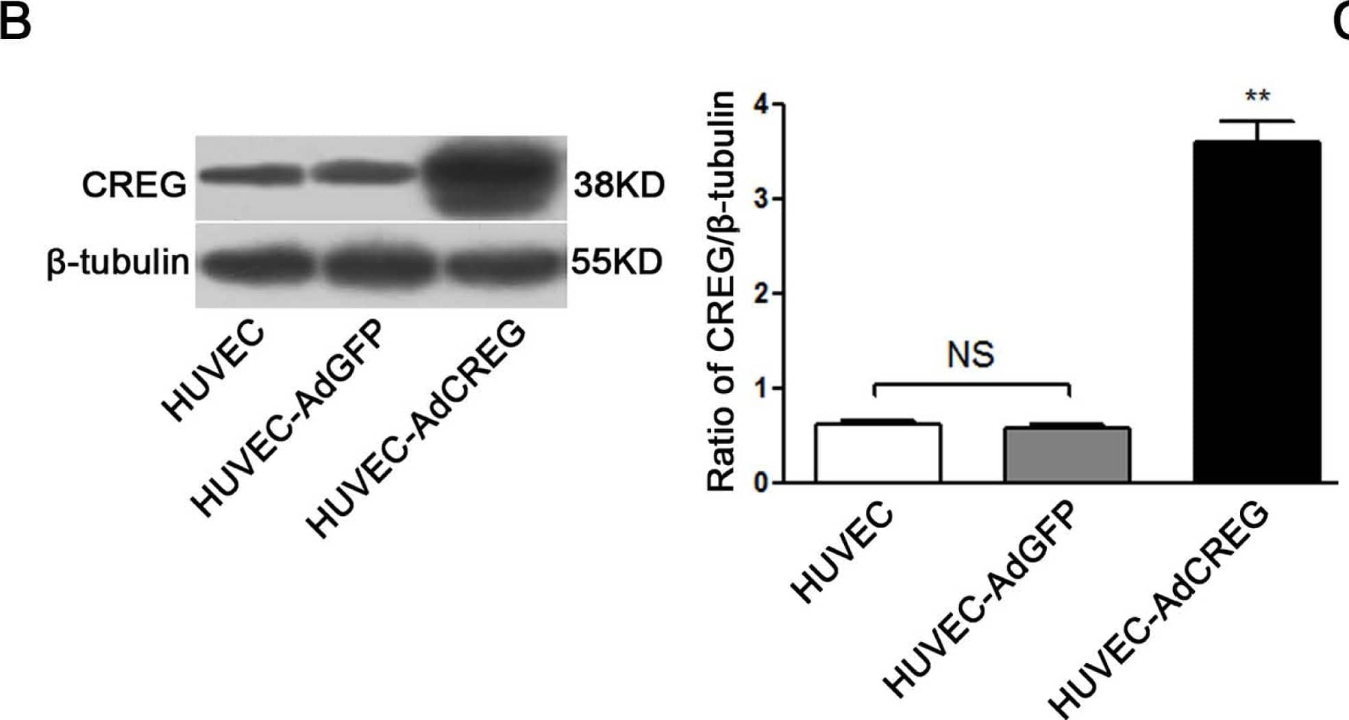
Overexpression of cellular repressor of E1A-stimulated genes (CREG) promotes the proliferation of human umbilical vein endothelial cells (HUVEC). (A) HUVECs infected with adenoviruses carrying CREG-IRES-GFP or GFP, respectively, have a similar infective efficiency. Images were taken by phase contrast microscope (a,c) and by fluorescence microscope (b,d) 48 h after infection. Bar = 25 μm; (B) CREG expression was detected by Western blotting. Levels of CREG were assessed after normalization to beta-tubulin. Data are given as the mean ± SD (n = 3). **p < 0.01 compared with uninfected HUVEC and HUVEC-expressing GFP (AdGFP) control groups. NS: no significant difference; (C) Growth curves of HUVECs, HUVEC-overexpressing CREG (AdCREG) and HUVEC-AdGFP were constructed by plotting cell numbers counted by hemocytometer over three days of incubation. **p < 0.01 compared with uninfected HUVEC and HUVEC-AdGFP control groups. Data are given as the mean ± SD (n = 3). (D) Flow cytometry (FCM) analysis of the cell cycle distributions of cells in three groups. Levels of proliferative potential were accessed by the percentage of cells in the (S + G2) phases of the cell cycle. ***p < 0.001 compared with HUVEC and HUVEC-AdGFP groups. NS: no significant difference. Data are given as the mean ± SD (n = 3). (E) 5-bromo-2′-deoxy-uridine (BrdU) incorporation assay of cells stained positively by diaminobenzidine (DAB) staining solution. The upper panel is a four-times high magnification image of the framed part in the lower panel. Levels of proliferation were assessed by the ratio of average BrdU positive cells to total cells in five random high-magnification fields. Data are given as the mean ± SD (n = 3). * p < 0.05 compared with HUVEC and HUVEC-AdGFP groups. NS: no significant difference. Bar = 25 μm. Image collected and cropped by CiteAb from the following open publication (https://pubmed.ncbi.nlm.nih.gov/24018888), licensed under a CC-BY license. Not internally tested by R&D Systems.Detection of Human CREG by Western Blot
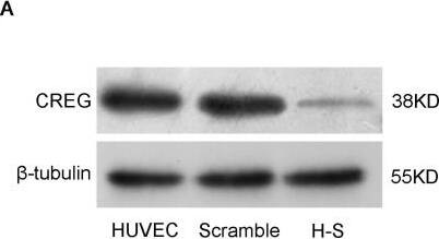
Downregulation of CREG suppresses the proliferation of HUVEC. (A) Stable HUVEC clones with CREG silenced down (H–S) or expressing a scrambled negative control shRNA sequence (scramble) were established by retroviral infection and puromycin selection. Cell lysates were collected, and the expression of CREG was detected by Western blotting with beta-tubulin used as a loading control; (B) Pooled analysis of CREG protein levels assessed after normalization to CREG/ beta-tubulin of the HUVEC group. Scramble HUVEC were used as a negative control. Data are given as the mean ± SD (n = 3). *p < 0.05; ***p < 0.001; NS: no significant difference; (C) Cell counting by hemocytometer. A total of 2 × 10⁵ cells in the logarithmic phase in each of the three groups were plated in four 10-cm diameter dishes. After 24 h, cells were trypsinized and counted by hemocytometer. **p < 0.01 compared with the HUVEC and scramble group; (D) BrdU incorporation assay. Data are given as the mean ± SD (n = 3). **p < 0.01; ***p < 0.001; NS: no significant difference; (E) Effects of the downregulation of CREG on HUVEC cell cycle progression assessed by FCM; (F) Cell proliferation was accessed by the percentage of (S + G2) phase cells in the cell cycle. Data are given as the mean ± SD (n = 3). **p < 0.01 compared with the HUVEC and scramble group. NS: no significant difference. Image collected and cropped by CiteAb from the following open publication (https://pubmed.ncbi.nlm.nih.gov/24018888), licensed under a CC-BY license. Not internally tested by R&D Systems.Applications for Human CREG Antibody
Application
Recommended Usage
Western Blot
1 µg/mL
Sample: Recombinant Human CREG
Sample: Recombinant Human CREG
Formulation, Preparation, and Storage
Purification
Protein A or G purified from hybridoma culture supernatant
Reconstitution
Reconstitute at 0.5 mg/mL in sterile PBS. For liquid material, refer to CoA for concentration.
Formulation
Lyophilized from a 0.2 μm filtered solution in PBS with Trehalose. *Small pack size (SP) is supplied either lyophilized or as a 0.2 µm filtered solution in PBS.
Shipping
Lyophilized product is shipped at ambient temperature. Liquid small pack size (-SP) is shipped with polar packs. Upon receipt, store immediately at the temperature recommended below.
Stability & Storage
Use a manual defrost freezer and avoid repeated freeze-thaw cycles.
- 12 months from date of receipt, -20 to -70 °C as supplied.
- 1 month, 2 to 8 °C under sterile conditions after reconstitution.
- 6 months, -20 to -70 °C under sterile conditions after reconstitution.
Background: CREG
References
- DiBacco, A. and G. Gill (2003) Oncogene 22:5436.
Long Name
Cellular Repressor of E1A-stimulated Genes
Alternate Names
CREG1
Gene Symbol
CREG1
UniProt
Additional CREG Products
Product Documents for Human CREG Antibody
Product Specific Notices for Human CREG Antibody
For research use only
Loading...
Loading...
Loading...
Loading...

