Human CX3CL1/Fractalkine Chemokine Domain Antibody
R&D Systems, part of Bio-Techne | Catalog # AF365


Key Product Details
Species Reactivity
Validated:
Cited:
Applications
Validated:
Cited:
Label
Antibody Source
Product Specifications
Immunogen
Gln25-Gly100
Accession # Q6I9S9
Specificity
Clonality
Host
Isotype
Endotoxin Level
Scientific Data Images for Human CX3CL1/Fractalkine Chemokine Domain Antibody
Chemotaxis Induced by CX3CL1/Fractalkine and Neutralization by Human CX3CL1/Fractalkine Antibody.
Recombinant Human CX3CL1/Fractalkine (Catalog # 365-FR) chemoattracts the BaF3 mouse pro-B cell line transfected with mouse CX3CR1 in a dose-dependent manner (orange line). The amount of cells that migrated through to the lower chemotaxis chamber was measured by Resazurin (Catalog # AR002). Chemotaxis elicited by Recombinant Human CX3CL1/Fractalkine (100 ng/mL) is neutralized (green line) by increasing concentrations of Goat Anti-Human CX3CL1/Fractalkine Chemokine Domain Antigen Affinity-purified Polyclonal Antibody (Catalog # AF365). The ND50 is typically 0.75-4.5 µg/mL.Detection of CX3CL1/Fractalkine by Western Blot
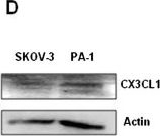
CX3CR1 is required for keratinocytes differentiation. (A) Expression of CX3CR1 in parental PA-1 cell line&that stably transfected with either CX3CR1 or scrambled shRNA constructs was examined with WB. Actin served as a loading control. CX3CR1 expression levels quantified using digital densitometry&normalized to the levels of actin expression. (B) Expression of cytokeratin 14 in PA-1 cells stably transfected with either CX3CR1 or scrambled shRNA constructs treated with recombinant BMP-4 or vehicle, as indicated, was examined with immunocytochemistry. CK14 in PA-1 was probed with anti-CK14&Alexa555-conjugated anti-rabbit antibodies,&DNA was stained with 4’,6-Diamidino-2-Phenylindole, Dihydrochloride (DAPI); CK14 – red, DNA – blue. Images taken using 5× magnification on the objective. The histogram demonstrates the average intensity of CK14 signal across the field as determined by the Zeiss AxioVision software. Data is an average of three independent experiments. *p < 0.05. (C) Expression of cytokeratins 14&18 in PA-1 cells stably transfected with either CX3CR1 or scrambled shRNA constructs treated with recombinant BMP-4 or vehicle, as indicated, was examined with WB. beta-Tubulin served as a loading control. The histogram shows CK14&CK18 expression levels. Expression of CK18 in CX3CR1shRNA-transfected PA-1 cells was arbitrarily set as 1&expression of both CK14&CK18 in other conditions was calculated accordingly. The data represent a typical WB image,&quantitative analysis was performed using three independently performed experiments. *p < 0.05. (D) Expression of CX3CL1 in SKOV-3&PA-1 cell lines was examined with WB. Actin served as a loading control. SKOV-3 cell line was used as a positive control. Image collected&cropped by CiteAb from the following open publication (https://pubmed.ncbi.nlm.nih.gov/23958497), licensed under a CC-BY license. Not internally tested by R&D Systems.Applications for Human CX3CL1/Fractalkine Chemokine Domain Antibody
Western Blot
Sample: Recombinant Human CX3CL1/Fractalkine Chemokine Domain (Catalog # 362-CX)
Neutralization
Reviewed Applications
Read 3 reviews rated 4.3 using AF365 in the following applications:
Formulation, Preparation, and Storage
Purification
Reconstitution
Formulation
Shipping
Stability & Storage
- 12 months from date of receipt, -20 to -70 °C as supplied.
- 1 month, 2 to 8 °C under sterile conditions after reconstitution.
- 6 months, -20 to -70 °C under sterile conditions after reconstitution.
Background: CX3CL1/Fractalkine
CX3CL1, also named neurotactin, is a novel chemokine identified through bioinformatics. CX3CL1 has a unique C-X3-C cysteine motif near the amino-terminus and is the first member of a fourth branch of the chemokine superfamily. Unlike other known chemokines, CX3CL1 is a type 1 membrane protein containing a chemokine domain tethered on a long mucin-like stalk. Human CX3CL1 cDNA encodes a 397 amino acid (aa) residue membrane protein with a 24 aa residue predicted signal peptide, a 76 aa residue chemokine domain, a 241 aa residue stalk region containing 17 degenerate mucin-like repeats, a 19 aa residue transmembrane segment and a 37 aa residue cytoplasmic domain. The extracellular domain of human CX3CL1 can be released, possibly by proteolysis at the dibasic cleavage site proximal to the membrane, to generate soluble CX3CL1. CX3CL1 mRNA has been detected in various tissues including the brain and heart. The expression of CX3CL1 was also reported to be up-regulated in endothelial cells and microglia by inflammatory signals. Membrane-bound CX3CL1 has been shown to promote adhesion of leukocytes. The soluble chemokine domain of human CX3CL1 was reported to be chemotactic for T cells and monocytes while the soluble chemokine domain of mouse CX3CL1 was reported to chemoattract neutrophils and T-lymphocytes but not monocytes. The gene for human CX3CL1 has been mapped to chromosome 16q.
References
- Pan, Y. et al. (1997) Nature 387:611.
- Bazan, J.F. et al. (1997) Nature 385:640.
- Mackay, C.R. (1997) Current Biology 7:R384.
Alternate Names
Gene Symbol
UniProt
Additional CX3CL1/Fractalkine Products
Product Documents for Human CX3CL1/Fractalkine Chemokine Domain Antibody
Product Specific Notices for Human CX3CL1/Fractalkine Chemokine Domain Antibody
For research use only
