Human CXCR4 Antibody
R&D Systems, part of Bio-Techne | Catalog # MAB171

Key Product Details
Validated by
Biological Validation
Species Reactivity
Validated:
Human
Cited:
Human, Xenograft
Applications
Validated:
Neutralization
Cited:
Binding Assay, FCCS, Flow Cytometry, Immunocytochemistry, Immunohistochemistry-Frozen, Immunohistochemistry-Paraffin, Neutralization, Western Blot
Label
Unconjugated
Antibody Source
Monoclonal Mouse IgG2A Clone # 44708
Product Specifications
Immunogen
3T3 cells transfected with human CXCR4
Specificity
Reacts specifically with human and non-human cells expressing human CXCR4 (fusin) as detected by flow cytometry. It will also react with cells expressing feline CXCR4 but not rat CXCR4. This antibody does not cross-react with other chemokine receptors.
Clonality
Monoclonal
Host
Mouse
Isotype
IgG2A
Endotoxin Level
<0.10 EU per 1 μg of the antibody by the LAL method.
Scientific Data Images for Human CXCR4 Antibody
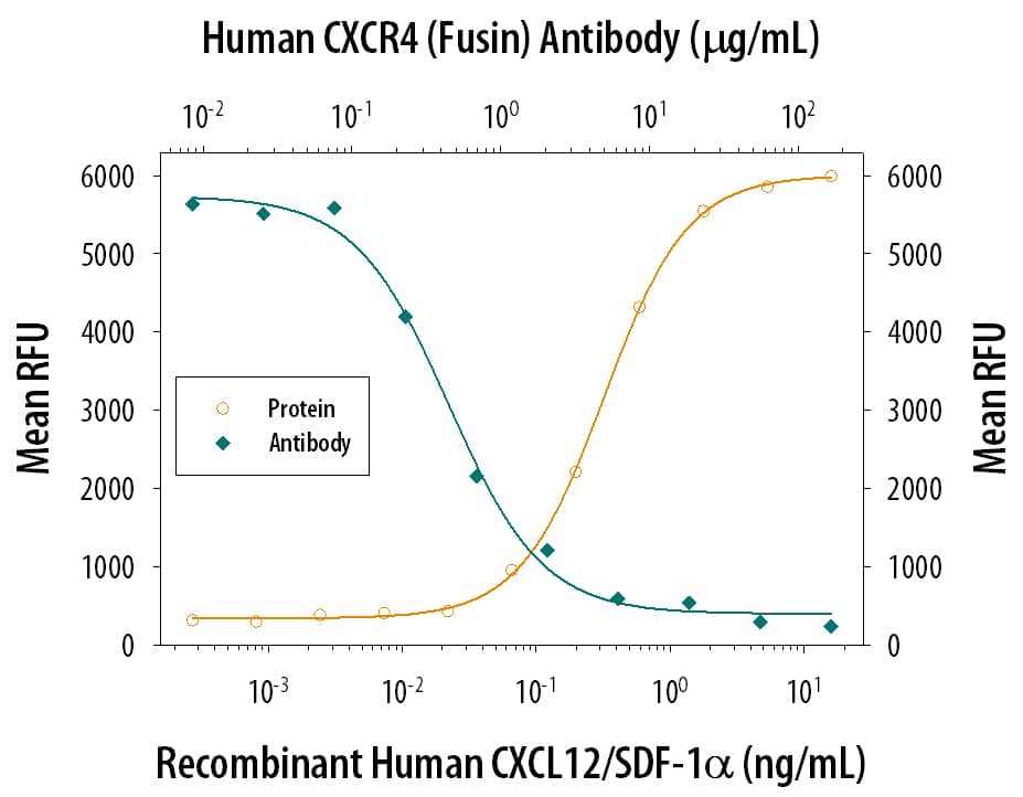
Chemotaxis Induced by CXCL12/SDF‑1 alpha and Neutralization by Human CXCR4 Antibody.
Recombinant Human/Feline/Rhesus Macaque CXCL12/SDF-1a (Catalog # 350-NS) chemoattracts the BaF3 mouse pro-B cell line transfected with human CXCR4 in a dose-dependent manner (orange line). The amount of cells that migrated through to the lower chemotaxis chamber was measured by Resazurin (Catalog # AR002). Chemotaxis elicited by Recombinant Human/Feline/Rhesus Macaque CXCL12/SDF-1a (1 ng/mL) is neutralized (green line) by increasing concentrations of Human CXCR4 Monoclonal Antibody (Catalog # MAB171). The ND50 is typically 0.2-1.2 µg/mL.Detection of Mouse CXCR4 by Flow Cytometry
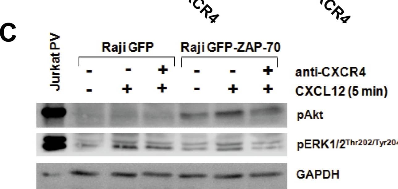
Anti-CXCR4 antibody reduces CXCR4 surface expression, and blocks migration toward CXCL12 and BMSCs.(A) Relative MFIR of CXCR4 surface staining after incubation of Raji transfectants with 100 µg/mL of anti-CXCR4 antibody for 30 minutes. (B) Flow cytometry histograms showing the reduction in CXCR4 expression after incubation with neutralizing anti-CXCR4 antibody. White histograms represent negative unstained controls. (C) CXCR4-blocked cells were stimulated with 100 ng/mL of CXCL12 for 5 minutes and activation of Akt and ERK1/2 proteins analyzed by western blotting. (D) CXCR4-blocked cells were subjected to migration assay for 4 hours at 37°C in 5% CO2 toward 100 ng/mL of CXCL12 (left panel) or the stromal MS-5 cell line (right panel). Cells in the lower chamber were counted by flow cytometry under a defined flow rate for 5 minutes. Number of migrated cells treated with isotypic control was considered 100%. Results are shown as the mean ± SEM of 4 independent experiments. (E) Cell viability was measured by Annexin V-PI staining in cells incubated with 100 µg/mL of anti-CXCR4 or isotypic control for 48 hours. Results are shown as the mean ± SEM of 4 independent experiments Image collected and cropped by CiteAb from the following publication (https://dx.plos.org/10.1371/journal.pone.0081221), licensed under a CC-BY license. Not internally tested by R&D Systems.Detection of Mouse CXCR4 by Western Blot
Anti-CXCR4 antibody reduces CXCR4 surface expression, and blocks migration toward CXCL12 and BMSCs.(A) Relative MFIR of CXCR4 surface staining after incubation of Raji transfectants with 100 µg/mL of anti-CXCR4 antibody for 30 minutes. (B) Flow cytometry histograms showing the reduction in CXCR4 expression after incubation with neutralizing anti-CXCR4 antibody. White histograms represent negative unstained controls. (C) CXCR4-blocked cells were stimulated with 100 ng/mL of CXCL12 for 5 minutes and activation of Akt and ERK1/2 proteins analyzed by western blotting. (D) CXCR4-blocked cells were subjected to migration assay for 4 hours at 37°C in 5% CO2 toward 100 ng/mL of CXCL12 (left panel) or the stromal MS-5 cell line (right panel). Cells in the lower chamber were counted by flow cytometry under a defined flow rate for 5 minutes. Number of migrated cells treated with isotypic control was considered 100%. Results are shown as the mean ± SEM of 4 independent experiments. (E) Cell viability was measured by Annexin V-PI staining in cells incubated with 100 µg/mL of anti-CXCR4 or isotypic control for 48 hours. Results are shown as the mean ± SEM of 4 independent experiments Image collected and cropped by CiteAb from the following publication (https://dx.plos.org/10.1371/journal.pone.0081221), licensed under a CC-BY license. Not internally tested by R&D Systems.Applications for Human CXCR4 Antibody
Application
Recommended Usage
Neutralization
Measured by its ability to neutralize CXCL12/SDF‑1 alpha-induced chemotaxis in the BaF3 mouse pro‑B cell line transfected with human CXCR4. The Neutralization Dose (ND50) is typically 0.2-1.2 µg/mL in the presence of 1 ng/mL Recombinant Human/Feline/Rhesus Macaque CXCL12/SDF‑1 alpha.
Formulation, Preparation, and Storage
Purification
Protein A or G purified from ascites
Reconstitution
Reconstitute at 0.5 mg/mL in sterile PBS. For liquid material, refer to CoA for concentration.
Formulation
Lyophilized from a 0.2 μm filtered solution in PBS with Trehalose. *Small pack size (SP) is supplied either lyophilized or as a 0.2 µm filtered solution in PBS.
Shipping
Lyophilized product is shipped at ambient temperature. Liquid small pack size (-SP) is shipped with polar packs. Upon receipt, store immediately at the temperature recommended below.
Stability & Storage
Use a manual defrost freezer and avoid repeated freeze-thaw cycles.
- 12 months from date of receipt, -20 to -70 °C as supplied.
- 1 month, 2 to 8 °C under sterile conditions after reconstitution.
- 6 months, -20 to -70 °C under sterile conditions after reconstitution.
Background: CXCR4
References
- Orsini, M.J. et al. (1999) J. Biol. Chem. 274:31076.
- Zagzag, D. et al. (2005) Cancer Res. 65:6178.
- Speetjens, F.M. et al. (2009) Cancer Microenvironment 2:1.
- Wang, L. et al. (2009) Oncology Reports 22:1333.
- Amara, S. et al. (2015) Cancer Biomark. 15:869.
Long Name
C-X-C Motif Chemokine Receptor 4
Alternate Names
CD184, D2S201E, FB22, Fusin, HM89, HSY3RR, LAP-3, LAP3, LCR1, LESTR, NPY3R, NPYRL, NPYY3R, WHIMS
Gene Symbol
CXCR4
Additional CXCR4 Products
Product Documents for Human CXCR4 Antibody
Product Specific Notices for Human CXCR4 Antibody
For research use only
Loading...
Loading...
Loading...
Loading...


