Human Cyclophilin A Antibody
R&D Systems, part of Bio-Techne | Catalog # MAB3589

Key Product Details
Species Reactivity
Validated:
Cited:
Applications
Validated:
Cited:
Label
Antibody Source
Product Specifications
Immunogen
Met1-Glu165
Accession # P62937
Specificity
Clonality
Host
Isotype
Scientific Data Images for Human Cyclophilin A Antibody
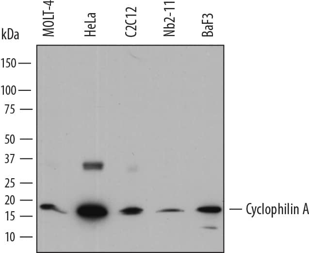
Detection of Human, Mouse, and Rat Cyclophilin A by Western Blot.
Western blot shows lysates of MOLT-4 human acute lymphoblastic leukemia cell line, HeLa human cervical epithelial carcinoma cell line, C2C12 mouse myoblast cell line, Nb2-11 rat lymphoma cell line, and BaF3 mouse pro-B cell line. PVDF membrane was probed with 0.1 µg/mL of Rat Anti-Human Cyclophilin A Monoclonal Antibody (Catalog # MAB3589) followed by HRP-conjugated Anti-Rat IgG Secondary Antibody (Catalog # HAF005). A specific band was detected for Cyclophilin A at approximately 18 kDa (as indicated). This experiment was conducted under reducing conditions and using Immunoblot Buffer Group 1.Cyclophilin A in PANC-1 Human Cell Line.
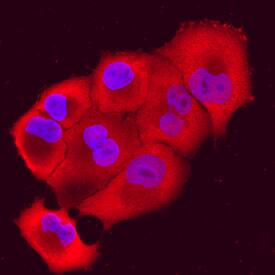
Cyclophilin A was detected in immersion fixed PANC-1 human pancreatic carcinoma cell line using Rat Anti-Human Cyclophilin A Monoclonal Antibody (Catalog # MAB3589) at 10 µg/mL for 3 hours at room temperature. Cells were stained using the NorthernLights™ 557-conjugated Anti-Rat IgG Secondary Antibody (red; Catalog # NL013) and counterstained with DAPI (blue). Specific staining was localized to nuclei and cytoplasm. View our protocol for Fluorescent ICC Staining of Cells on Coverslips.Detection of Human Cyclophilin A by Simple WesternTM.
Simple Western shows lysates of MOLT-4 human acute lymphoblastic leukemia cell line, loaded at 0.5 mg/ml. A specific band was detected for Cyclophilin A at approximately 22 kDa (as indicated) using 10 µg/mL of Rat Anti-Human Cyclophilin A Monoclonal Antibody (Catalog # MAB3589). This experiment was conducted under reducing conditions and using the 2-40kDa separation system.Applications for Human Cyclophilin A Antibody
Immunocytochemistry
Sample: Immersion fixed PANC1 human pancreatic carcinoma cell line
Simple Western
Sample: MOLT-4 human acute lymphoblastic leukemia cell line
Western Blot
Sample: MOLT‑4 human acute lymphoblastic leukemia cell line, HeLa human cervical epithelial carcinoma cell line, C2C12 mouse myoblast cell line, Nb2‑11 rat lymphoma cell line, and BaF3 mouse pro-B cell line
Reviewed Applications
Read 1 review rated 5 using MAB3589 in the following applications:
Formulation, Preparation, and Storage
Purification
Reconstitution
Formulation
*Small pack size (-SP) is supplied either lyophilized or as a 0.2 µm filtered solution in PBS.
Shipping
Stability & Storage
- 12 months from date of receipt, -20 to -70 °C as supplied.
- 1 month, 2 to 8 °C under sterile conditions after reconstitution.
- 6 months, -20 to -70 °C under sterile conditions after reconstitution.
Background: Cyclophilin A
Cyclophilin A, also called Peptidyl-prolyl Isomerase A, PPIA, CYPA, and CYPH, was originally characterized for its ability to catalyze the transition between cis- and trans- proline residues critical for proper folding of proteins (1). Cyclophilin is also incorporated into many viruses, including HIV-1, where it has been speculated to be involved in functions such as viral assembly and infectivity (2). The immunosuppressive activity of cyclosporins has been correlated with their ability to form complexes with cyclophilins that inhibit calcineurin phosphatase activity (3) and prevent incorporation of cyclophilin into viral particles (4). The cyclosporin/cyclophilin complex selectively binds and inactivates calcineurin (3, 5), making it a useful inhibitor for studying calcineurin activity.
References
- Hamilton, G.S. and J.P. Steiner (1998) J. Med. Chem. 41:5119.
- Cantin, R. et al. (2005) J. Virology 79:6577.
- Liu, J. et al. (1992) Biochemistry 31:3896.
- Wiegers K. and H.G. Krausslich (2002) Virology 294:289.
- Liu, J. et al. (1991) Cell 66:807.
Alternate Names
Gene Symbol
UniProt
Additional Cyclophilin A Products
Product Documents for Human Cyclophilin A Antibody
Product Specific Notices for Human Cyclophilin A Antibody
For research use only


