Human DEP-1/CD148 Antibody
R&D Systems, part of Bio-Techne | Catalog # MAB1934


Key Product Details
Species Reactivity
Validated:
Cited:
Applications
Validated:
Cited:
Label
Antibody Source
Product Specifications
Immunogen
Specificity
Clonality
Host
Isotype
Scientific Data Images for Human DEP-1/CD148 Antibody
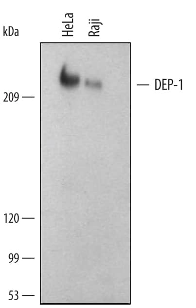
Detection of Human DEP-1/CD148 by Western Blot.
Western blot shows lysates of HeLa human cervical epithelial carcinoma cell line and Raji human Burkitt's lymphoma cell line. PVDF membrane was probed with 1 µg/mL of Human DEP-1/CD148 Monoclonal Antibody (Catalog # MAB1934) followed by HRP-conjugated Anti-Mouse IgG Secondary Antibody (Catalog # HAF007). A specific band was detected for DEP-1/CD148 at approximately 220 kDa (as indicated). This experiment was conducted under reducing conditions and using Immunoblot Buffer Group 1.Applications for Human DEP-1/CD148 Antibody
CyTOF-ready
Flow Cytometry
Sample: Human peripheral blood lymphocytes
Immunoprecipitation
Sample: HeLa human cervical epithelial carcinoma cell line, see our available Western blot detection antibodies
Western Blot
Sample: HeLa human cervical epithelial carcinoma cell line and Raji human Burkitt's lymphoma cell line
Reviewed Applications
Read 2 reviews rated 4.5 using MAB1934 in the following applications:
Formulation, Preparation, and Storage
Purification
Reconstitution
Formulation
Shipping
Stability & Storage
- 12 months from date of receipt, -20 to -70 °C as supplied.
- 1 month, 2 to 8 °C under sterile conditions after reconstitution.
- 6 months, -20 to -70 °C under sterile conditions after reconstitution.
Background: DEP-1/CD148
Density Enhanced Protein Tyrosine Phosphatase (DEP-1), also known as CD148, HPTP-eta, and PTP receptor type J (PTPRJ), is an enzyme that removes phosphate groups covalently attached to tyrosine residues in proteins. A large (220 kDa) glycoprotein found at the cell surface, DEP-1 levels are increased with high cell density (1). DEP-1 phosphatase activity is enhanced by basement membrane proteins (2), suggesting it is involved in regulating cell adhesion and contact interactions. High levels of expression dampen PDGF (3), VEGF (4), and T‑cell receptor (5) responses. DEP-1 is widely expressed in tissues, particularly ones forming epithelioid monolayers (6). In the immune system, DEP-1 is found on all cell lineages and is highest on granulocytes (7). Dep-1 is the mutated gene in the Susceptibility to Colon Cancer locus Scc1, which is altered in many human colorectal adenomas (8). Gene knockout mice lacking DEP-1 die at midgestation due to failures in cardiovascular development (9). DEP-1 dephosphorylates a variety of proteins, including the HGF (10), PDGF (11), and VEGF (4) receptors, and beta-catenin (12). The recombinant protein is the intracellular region of DEP-1 containing the catalytic domain.
References
- Ostman, A. et al. (1994) Proc. Natl. Acad. Sci. USA 91:9680.
- Sorby, M. et al. (2001) Oncogene 20:5219.
- Jandt, E. et al. (2003) Oncogene 22:4175.
- Lampugnani, M.G. et al. (2003) J. Cell Biol. 161:793.
- Baker, J.E. et al. (2001) Mol. Cell. Biol. 21:2393.
- Borges, L.G. et al. (1996) Circ. Res. 79:570.
- de la Fuente-Garcia, M.A. et al. (1998) Blood 91:2800.
- Ruivenkamp, C.A. et al. (2002) Nat. Genet. 31:295.
- Takahashi, T. et al. (2003) Mol. Cell. Biol. 23:1817.
- Palka, H.L. et al. (2003) J. Biol. Chem. 278:5728.
- Kovalenko, M. et al. (2000) J. Biol. Chem. 275:16219.
- Holsinger, L.J. et al. (2002) Oncogene 21:7067.
Long Name
Alternate Names
Gene Symbol
Additional DEP-1/CD148 Products
Product Documents for Human DEP-1/CD148 Antibody
Product Specific Notices for Human DEP-1/CD148 Antibody
For research use only