Human Dihydrofolate Reductase/DHFR Antibody
R&D Systems, part of Bio-Techne | Catalog # AF7934

Key Product Details
Species Reactivity
Applications
Label
Antibody Source
Product Specifications
Immunogen
Met1-Asp187
Accession # P00374
Specificity
Clonality
Host
Isotype
Scientific Data Images for Human Dihydrofolate Reductase/DHFR Antibody
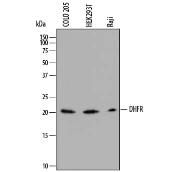
Detection of Human Dihydrofolate Reductase/DHFR by Western Blot.
Western blot shows lysates of COLO 205 human colorectal adenocarcinoma cell line, HEK293T human embryonic kidney cell line, and Raji human Burkitt's lymphoma cell line. PVDF membrane was probed with 0.5 µg/mL of Sheep Anti-Human Dihydrofolate Reductase/DHFR Antigen Affinity-purified Polyclonal Antibody (Catalog # AF7934) followed by HRP-conjugated Anti-Sheep IgG Secondary Antibody (Catalog # HAF016). A specific band was detected for Dihydrofolate Reductase/DHFR at approximately 21 kDa (as indicated). This experiment was conducted under reducing conditions and using Immunoblot Buffer Group 1.Dihydrofolate Reductase/DHFR in MCF‑7 Human Cell Line.
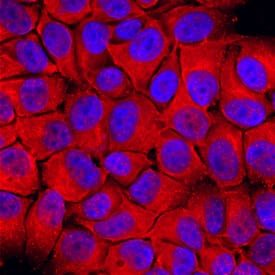
Dihydrofolate Reductase/ DHFR was detected in immersion fixed MCF-7 human breast cancer cell line using Sheep Anti-Human Dihydrofolate Reductase/ DHFR Antigen Affinity-purified Polyclonal Antibody (Catalog # AF7934) at 10 µg/mL for 3 hours at room temperature. Cells were stained using the Northern-Lights™ 557-conjugated Anti-Sheep IgG Secondary Antibody (red; Catalog # NL010) and counter-stained with DAPI (blue). Specific staining was localized to cytoplasm. View our protocol for Fluorescent ICC Staining of Cells on Coverslips.Applications for Human Dihydrofolate Reductase/DHFR Antibody
Immunocytochemistry
Sample: Immersion fixed MCF‑7 human breast cancer cell line
Western Blot
Sample: COLO 205 human colorectal adenocarcinoma cell line, HEK293T human embryonic kidney cell line, and Raji human Burkitt's lymphoma cell line
Formulation, Preparation, and Storage
Purification
Reconstitution
Formulation
Shipping
Stability & Storage
- 12 months from date of receipt, -20 to -70 °C as supplied.
- 1 month, 2 to 8 °C under sterile conditions after reconstitution.
- 6 months, -20 to -70 °C under sterile conditions after reconstitution.
Background: Dihydrofolate Reductase/DHFR
DHFR (DiHydroFolate Reductase; also Tetrahydrofolate dehydrogenase) is a 21-23 kDa member of the dihydrofolate reductase family of enzymes. It is a ubiquitously expressed monomer, and considered to be a housekeeping gene. Housekeeping genes are those that play a role in multiple pathways, although not the same pathway(s) in all cells. DHFR participates in the reduction of dihydrofolate to tetrahydrofolate, a product that is subsequently used in the synthesis of purines and thymidylic acid that are used to generate both RNA and DNA. Within the cell, DHFR is known to exist in two pools: one contains DHFR bound to its own RNA where it acts as a transcriptional repressor, while another contains DHFR bound to NADPH. Human DHFR is 187 amino acids (aa) in length and possesses one DHFR domain (aa 4-185). Its mRNA binding motif is suggested to involve Cys6, Leu22, Glu30 and Ser118. There is one potential alternative start site found 75 aa upstream of the standard start site. Full length human DHFR (aa 1-187) shares 90% aa sequence identity with mouse DHFR.
Alternate Names
Gene Symbol
UniProt
Additional Dihydrofolate Reductase/DHFR Products
Product Documents for Human Dihydrofolate Reductase/DHFR Antibody
Product Specific Notices for Human Dihydrofolate Reductase/DHFR Antibody
For research use only

