Human DPP9 Antibody
R&D Systems, part of Bio-Techne | Catalog # MAB54191

Key Product Details
Species Reactivity
Applications
Label
Antibody Source
Product Specifications
Immunogen
Arg2-Leu892
Accession # Q86TI2
Specificity
Clonality
Host
Isotype
Scientific Data Images for Human DPP9 Antibody
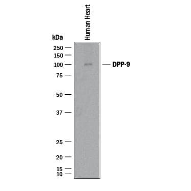
Detection of Human DPP9 by Western Blot.
Western blot shows lysates of human heart tissue. PVDF membrane was probed with 1 µg/mL of Mouse Anti-Human DPP9 Monoclonal Antibody (Catalog # MAB54191) followed by HRP-conjugated Anti-Mouse IgG Secondary Antibody (Catalog # HAF018). A specific band was detected for DPP9 at approximately 100 kDa (as indicated). This experiment was conducted under reducing conditions and using Immunoblot Buffer Group 1.DPP9 in HeLa Human Cell Line.
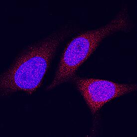
DPP9 was detected in immersion fixed HeLa human cervical epithelial carcinoma cell line using Mouse Anti-Human DPP9 Monoclonal Antibody (Catalog # MAB54191) at 8 µg/mL for 3 hours at room temperature. Cells were stained using the NorthernLights™ 557-conjugated Anti-Mouse IgG Secondary Antibody (red; Catalog # NL007) and counterstained with DAPI (blue). Specific staining was localized to cytoplasm and nuclei. View our protocol for Fluorescent ICC Staining of Cells on Coverslips.Detection of Human DPP9 by Simple WesternTM.
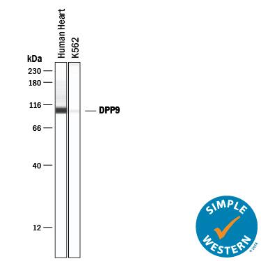
Simple Western lane view shows lysates of human heart tissue and K562 human chronic myelogenous leukemia cell line, loaded at 0.2 mg/mL. A specific band was detected for DPP9 at approximately 104 kDa (as indicated) using 10 µg/mL of Mouse Anti-Human DPP9 Monoclonal Antibody (Catalog # MAB54191) . This experiment was conducted under reducing conditions and using the 12-230 kDa separation system.Applications for Human DPP9 Antibody
Immunocytochemistry
Sample: Immersion fixed HeLa human cervical epithelial carcinoma cell line
Simple Western
Sample: Human heart tissue and K562 human chronic myelogenous leukemia cell line
Western Blot
Sample: Human heart tissue
Formulation, Preparation, and Storage
Purification
Reconstitution
Formulation
Shipping
Stability & Storage
- 12 months from date of receipt, -20 to -70 °C as supplied.
- 1 month, 2 to 8 °C under sterile conditions after reconstitution.
- 6 months, -20 to -70 °C under sterile conditions after reconstitution.
Background: DPP9
DPP9 is a member of the S9b family of serine peptidases (1, 2). It shares 19% amino acid identity with DPP4 and 58% amino acid identity with DPP8. It exhibits post‑proline dipeptidyl aminopeptidase activity, cleaving Xaa-Pro dipeptides from the N-terminus of oligo- and polypeptides (3). Unlike DPP4, DPP9 does not appear to be membrane bound and is localized exclusively in the cytoplasm (4). This family of proline-specific dipeptidyl peptidases has been implicated in a variety of diseases including type 2 diabetes, obesity and cancer, and has been a potential target for drug discovery (5, 6).
References
- Olsen, C. and Wagtmann, N. (2002) Gene 299:185.
- Qi, S.Y. et al. (2003) Biochem. J. 373:179.
- Bjelke, J.R. et al. (2006) Biochem. J. 396:391.
- Ajami, K. et al. (2004) Biochim. Biophys. Acta. 1679:18.
- Rosenblum, J.S. and Kozarich, J.W. et al. (2003) Curr. Opin. Chem. Biol. 7:496.
- Van der Veken, P. et al. (2007) Curr. Top. Med. Chem. 7:621.
Long Name
Alternate Names
Gene Symbol
UniProt
Additional DPP9 Products
Product Documents for Human DPP9 Antibody
Product Specific Notices for Human DPP9 Antibody
For research use only


